| Entry | Database: PDB / ID: 3lrc
|
|---|
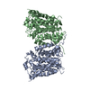
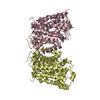
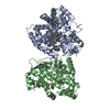
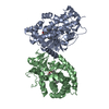
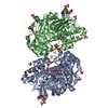
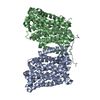
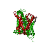
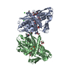
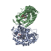
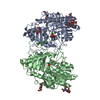
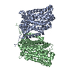
| Title | Structure of E. coli AdiC (P1) |
|---|
 Components Components | Arginine/agmatine antiporter |
|---|
 Keywords Keywords |  TRANSPORT PROTEIN / AdiC / transporter / antiporter / Amino-acid transport / Antiport / Cell inner membrane / Cell membrane / Membrane / Transmembrane / Transport TRANSPORT PROTEIN / AdiC / transporter / antiporter / Amino-acid transport / Antiport / Cell inner membrane / Cell membrane / Membrane / Transmembrane / Transport |
|---|
| Function / homology | Amino acid/polyamine transporter I / Amino acid permease / amino acid transport / antiporter activity / identical protein binding / plasma membrane / Arginine/agmatine antiporter Function and homology information Function and homology information |
|---|
| Biological species |   Escherichia coli (E. coli) Escherichia coli (E. coli) |
|---|
| Method | X-RAY DIFFRACTION / SYNCHROTRON / MOLECULAR REPLACEMENT / Resolution: 4.004 Å |
|---|
 Authors Authors | Gao, X. / Lu, F. / Zhou, L. / Shi, Y. |
|---|
 Citation Citation | Journal: Science / Year: 2009
Title: Structure and mechanism of an amino acid antiporter
Authors: Gao, X. / Lu, F. / Zhou, L. / Dang, S. / Sun, L. / Li, X. / Wang, J. / Shi, Y. |
|---|
| History | | Deposition | Feb 11, 2010 | Deposition site: RCSB / Processing site: PDBJ |
|---|
| Supersession | Feb 23, 2010 | ID: 3H6B |
|---|
| Revision 1.0 | Feb 23, 2010 | Provider: repository / Type: Initial release |
|---|
| Revision 1.1 | Jul 13, 2011 | Group: Version format compliance |
|---|
| Revision 1.2 | Nov 1, 2023 | Group: Data collection / Database references / Refinement description
Category: chem_comp_atom / chem_comp_bond ...chem_comp_atom / chem_comp_bond / database_2 / pdbx_initial_refinement_model / struct_ncs_dom_lim
Item: _database_2.pdbx_DOI / _database_2.pdbx_database_accession ..._database_2.pdbx_DOI / _database_2.pdbx_database_accession / _struct_ncs_dom_lim.beg_auth_comp_id / _struct_ncs_dom_lim.beg_label_asym_id / _struct_ncs_dom_lim.beg_label_comp_id / _struct_ncs_dom_lim.beg_label_seq_id / _struct_ncs_dom_lim.end_auth_comp_id / _struct_ncs_dom_lim.end_label_asym_id / _struct_ncs_dom_lim.end_label_comp_id / _struct_ncs_dom_lim.end_label_seq_id |
|---|
|
|---|
 Movie
Movie Controller


 Open data
 Basic information
 Structure visualization
 Downloads & links
 Other downloads


 PDBj
PDBj Assembly