 Movie
Movie Controller


+ Open data

- Basic information
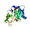
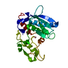
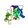
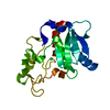
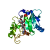
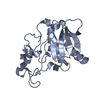
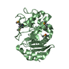
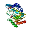
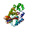
| Entry | Database: PDB / ID: 3lq0 | ||||||
|---|---|---|---|---|---|---|---|
| Title | Zymogen structure of crayfish astacin metallopeptidase | ||||||
 Components Components | ProAstacin | ||||||
 Keywords Keywords |  HYDROLASE / metallopeptidase / zymogen activation / proenzyme / protease / Disulfide bond / Metal-binding / Metalloprotease / Zymogen HYDROLASE / metallopeptidase / zymogen activation / proenzyme / protease / Disulfide bond / Metal-binding / Metalloprotease / Zymogen | ||||||
| Function / homology |  Function and homology informationastacin / glutamic-type peptidase activity / negative regulation of binding of sperm to zona pellucida / aspartic-type peptidase activity / prevention of polyspermy / cortical granule / positive regulation of protein processing / fertilization / metalloendopeptidase activity / peptidase activity ...astacin / glutamic-type peptidase activity / negative regulation of binding of sperm to zona pellucida / aspartic-type peptidase activity / prevention of polyspermy / cortical granule / positive regulation of protein processing / fertilization / metalloendopeptidase activity / peptidase activity / cell adhesion / proteolysis / zinc ion binding / plasma membrane / cytoplasm Function and homology informationastacin / glutamic-type peptidase activity / negative regulation of binding of sperm to zona pellucida / aspartic-type peptidase activity / prevention of polyspermy / cortical granule / positive regulation of protein processing / fertilization / metalloendopeptidase activity / peptidase activity ...astacin / glutamic-type peptidase activity / negative regulation of binding of sperm to zona pellucida / aspartic-type peptidase activity / prevention of polyspermy / cortical granule / positive regulation of protein processing / fertilization / metalloendopeptidase activity / peptidase activity / cell adhesion / proteolysis / zinc ion binding / plasma membrane / cytoplasmSimilarity search - Function | ||||||
| Biological species |  Astacus astacus (noble crayfish) Astacus astacus (noble crayfish) | ||||||
| Method | X-RAY DIFFRACTION / SYNCHROTRON / MOLECULAR REPLACEMENT / Resolution: 1.45 Å | ||||||
 Authors Authors | Guevara, T. / Yiallouros, I. / Kappelhoff, R. / Bissdorf, S. / Stocker, W. / Gomis-Ruth, F.X. | ||||||
 Citation Citation | Journal: J.Biol.Chem. / Year: 2010 Title: Proenzyme structure and activation of astacin metallopeptidase Authors: Guevara, T. / Yiallouros, I. / Kappelhoff, R. / Bissdorf, S. / Stocker, W. / Gomis-Ruth, F.X. | ||||||
| History |
|
- Structure visualization
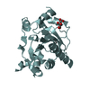
| Structure viewer | Molecule: MolmilJmol/JSmol |
|---|
- Downloads & links
-Download
| PDBx/mmCIF format | 3lq0.cif.gz | 125.4 KB | Display | PDBx/mmCIF format |
|---|---|---|---|---|
| PDB format | pdb3lq0.ent.gz | 96.1 KB | Display | PDB format |
| PDBx/mmJSON format | 3lq0.json.gz | Tree view | PDBx/mmJSON format | |
| Others |  Other downloads Other downloads |
-Validation report
| Arichive directory | https://data.pdbj.org/pub/pdb/validation_reports/lq/3lq0ftp://data.pdbj.org/pub/pdb/validation_reports/lq/3lq0 | HTTPS FTP |
|---|
-Related structure data
| Related structure data |  1astS S: Starting model for refinement |
|---|---|
| Similar structure data |


-Links
 PDBj
PDBj


- Assembly
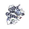
| Deposited unit | 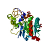
| ||||||||
|---|---|---|---|---|---|---|---|---|---|
| 1 |
| ||||||||
| Unit cell |
|
-Components
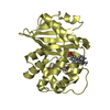
| #1: Protein | Mass: 26437.385 Da / Num. of mol.: 1 / Mutation: I91L,E93A Source method: isolated from a genetically manipulated source Details: Recombinant proastacin Glu93MAla, Ile91MLeu (UniProt Q9U918; numbering is based on the mature enzyme, see below) was expressed in Escherichia coli BL21(DE3) cells as inclusion bodies, ...Details: Recombinant proastacin Glu93MAla, Ile91MLeu (UniProt Q9U918; numbering is based on the mature enzyme, see below) was expressed in Escherichia coli BL21(DE3) cells as inclusion bodies, purified by Ni-NTA-affinity chromatography, and folded by dilution and removal of reducing agents and guanidinium chloride. Source: (gene. exp.) Astacus astacus (noble crayfish) / Plasmid: pET3a / Production host:  Escherichia coli (E. coli) / Strain (production host): BL21(DE3) / References: UniProt: P07584, astacin Escherichia coli (E. coli) / Strain (production host): BL21(DE3) / References: UniProt: P07584, astacin | ||||||
|---|---|---|---|---|---|---|---|
| #2: Chemical | ChemComp-ZN /   Mass: 65.409 Da / Num. of mol.: 1 / Source method: obtained synthetically / Formula: Zn Mass: 65.409 Da / Num. of mol.: 1 / Source method: obtained synthetically / Formula: Zn | ||||||
| #3: Chemical | Glycerol  Mass: 92.094 Da / Num. of mol.: 3 / Source method: obtained synthetically / Formula: C3H8O3 Mass: 92.094 Da / Num. of mol.: 3 / Source method: obtained synthetically / Formula: C3H8O3#4: Chemical | ChemComp-SO4 / | Sulfate  Mass: 96.063 Da / Num. of mol.: 1 / Source method: obtained synthetically / Formula: SO4 Mass: 96.063 Da / Num. of mol.: 1 / Source method: obtained synthetically / Formula: SO4#5: Water | ChemComp-HOH / | Water Mass: 18.015 Da / Num. of mol.: 298 / Source method: isolated from a natural source / Formula: H2O Mass: 18.015 Da / Num. of mol.: 298 / Source method: isolated from a natural source / Formula: H2OCompound details | THE FIRST 34 RESIDUES ARE FOR ACTIVATION | |
-Experimental details
-Experiment
| Experiment | Method: X-RAY DIFFRACTION / Number of used crystals: 1 |
|---|
- Sample preparation
| Crystal | Density Matthews: 2.32 Å3/Da / Density % sol: 46.94 % |
|---|---|
| Crystal grow | Temperature: 277 K / Method: vapor diffusion, sitting drop / pH: 5.6 Details: For crystallization, reservoir solutions were prepared by a Tecan robot and 200-nL crystallization drops were dispensed on 96x2-well MRC plates (Innovadyne) by a Cartesian (Genomic Solutions) ...Details: For crystallization, reservoir solutions were prepared by a Tecan robot and 200-nL crystallization drops were dispensed on 96x2-well MRC plates (Innovadyne) by a Cartesian (Genomic Solutions) nanodrop robot at the High-Throughput Crystallography Platform of the Barcelona Science Park. Best crystals appeared in a Bruker steady-temperature crystal farm at 4C with protein solution (10 mg/mL in 50mM AMPSO pH9.0) and 20% PEG 8000, 0.1M (NH4)2SO4, 0.01M MgCl2, 0.05M MES pH5.6 as reservoir solution. These conditions were efficiently scaled up to the microliter range with 24-well Cryschem crystallization dishes (Hampton Research). Crystals were cryo-protected with 16% PEG 8000, 20% glycerol, 0.1M (NH4)2SO4, 0.01M MgCl2, 0.05M MES pH5.6. , VAPOR DIFFUSION, SITTING DROP, temperature 277K |
-Data collection
| Diffraction | Mean temperature: 100 K |
|---|---|
| Diffraction source | Source: SYNCHROTRON / Site: ESRF  / Beamline: ID23-2 / Wavelength: 0.873 Å / Beamline: ID23-2 / Wavelength: 0.873 Å |
| Detector | Type: MAR CCD 165 mm / Detector: CCD / Date: Oct 6, 2009 |
| Radiation | Monochromator: horizontally diffracting Si (111) monochromator Protocol: SINGLE WAVELENGTH / Monochromatic (M) / Laue (L): M / Scattering type: x-ray |
| Radiation wavelength | Wavelength: 0.873 Å / Relative weight: 1 |
| Reflection | Resolution: 1.45→46.73 Å / Num. all: 44316 / Num. obs: 44227 / % possible obs: 99.8 % / Observed criterion σ(F): 1.44 / Observed criterion σ(I): 2 / Redundancy: 7.1 % / Biso Wilson estimate: 14.6 Å2 / Rmerge(I) obs: 0.044 / Rsym value: 0.044 / Net I/σ(I): 27.3 |
| Reflection shell | Resolution: 1.45→1.54 Å / Redundancy: 6.2 % / Rmerge(I) obs: 0.474 / Mean I/σ(I) obs: 3.8 / Num. unique all: 6285 / Rsym value: 0.474 / % possible all: 98.9 |
- Processing
| Software |
| ||||||||||||||||||||||||||||||||||||||||||||||||||||||||||||||||||||||||||||||||||||||||||||||||||||
|---|---|---|---|---|---|---|---|---|---|---|---|---|---|---|---|---|---|---|---|---|---|---|---|---|---|---|---|---|---|---|---|---|---|---|---|---|---|---|---|---|---|---|---|---|---|---|---|---|---|---|---|---|---|---|---|---|---|---|---|---|---|---|---|---|---|---|---|---|---|---|---|---|---|---|---|---|---|---|---|---|---|---|---|---|---|---|---|---|---|---|---|---|---|---|---|---|---|---|---|---|---|
| Refinement | Method to determine structure: MOLECULAR REPLACEMENT Starting model: PDB ENTRY 1AST Resolution: 1.45→41.92 Å / Cor.coef. Fo:Fc: 0.972 / Cor.coef. Fo:Fc free: 0.957 / SU B: 2.29 / SU ML: 0.039 / Cross valid method: THROUGHOUT / σ(F): 0 / σ(I): 0 / ESU R: 0.069 / ESU R Free: 0.06 / Stereochemistry target values: MAXIMUM LIKELIHOOD Details: HYDROGENS HAVE BEEN ADDED IN THE RIDING POSITIONS.; THE NUMBERING USED IN THE PRIMARY CITATION IS: RESIDUES 1 TO 34 AND CHAIN P FOR THE PROPEPTIDE AND RESIDUES 1 TO 201 AND CHAIN M FOR THE ...Details: HYDROGENS HAVE BEEN ADDED IN THE RIDING POSITIONS.; THE NUMBERING USED IN THE PRIMARY CITATION IS: RESIDUES 1 TO 34 AND CHAIN P FOR THE PROPEPTIDE AND RESIDUES 1 TO 201 AND CHAIN M FOR THE MATURE PROTEASE MOIETY.
| ||||||||||||||||||||||||||||||||||||||||||||||||||||||||||||||||||||||||||||||||||||||||||||||||||||
| Solvent computation | Ion probe radii: 0.8 Å / Shrinkage radii: 0.8 Å / VDW probe radii: 1.4 Å / Solvent model: BABINET MODEL WITH MASK | ||||||||||||||||||||||||||||||||||||||||||||||||||||||||||||||||||||||||||||||||||||||||||||||||||||
| Displacement parameters | Biso mean: 7.029 Å2
| ||||||||||||||||||||||||||||||||||||||||||||||||||||||||||||||||||||||||||||||||||||||||||||||||||||
| Refinement step | Cycle: LAST / Resolution: 1.45→41.92 Å
| ||||||||||||||||||||||||||||||||||||||||||||||||||||||||||||||||||||||||||||||||||||||||||||||||||||
| Refine LS restraints |
| ||||||||||||||||||||||||||||||||||||||||||||||||||||||||||||||||||||||||||||||||||||||||||||||||||||
| LS refinement shell | Resolution: 1.45→1.488 Å / Total num. of bins used: 20
| ||||||||||||||||||||||||||||||||||||||||||||||||||||||||||||||||||||||||||||||||||||||||||||||||||||
| Refinement TLS params. | Method: refined / Refine-ID: X-RAY DIFFRACTION
| ||||||||||||||||||||||||||||||||||||||||||||||||||||||||||||||||||||||||||||||||||||||||||||||||||||
| Refinement TLS group |
|
