 Movie
Movie Controller


+ Open data

- Basic information
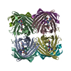
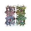
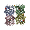
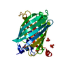
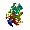
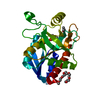
| Entry | Database: PDB / ID: 3p8u | |||||||||
|---|---|---|---|---|---|---|---|---|---|---|
| Title | Crystal structure of mEosFP in its green state | |||||||||
 Components Components | Green to red photoconvertible GPF-like protein EosFP | |||||||||
 Keywords Keywords |  FLUORESCENT PROTEIN / Beta-barrel FLUORESCENT PROTEIN / Beta-barrel | |||||||||
| Function / homology |  Function and homology information Function and homology information | |||||||||
| Biological species |  Lobophyllia hemprichii (invertebrata) Lobophyllia hemprichii (invertebrata) | |||||||||
| Method | X-RAY DIFFRACTION / SYNCHROTRON / MOLECULAR REPLACEMENT / Resolution: 2.25 Å | |||||||||
 Authors Authors | Adam, V. / Nienhaus, G.U. / Bourgeois, D. | |||||||||
 Citation Citation | Journal: Chem.Biol. / Year: 2011 Title: Rational design of photoconvertible and biphotochromic fluorescent proteins for advanced microscopy applications. Authors: Adam, V. / Moeyaert, B. / David, C.C. / Mizuno, H. / Lelimousin, M. / Dedecker, P. / Ando, R. / Miyawaki, A. / Michiels, J. / Engelborghs, Y. / Hofkens, J. | |||||||||
| History |
|
- Structure visualization
| Structure viewer | Molecule: MolmilJmol/JSmol |
|---|
- Downloads & links
-Download
| PDBx/mmCIF format | 3p8u.cif.gz | 200.3 KB | Display | PDBx/mmCIF format |
|---|---|---|---|---|
| PDB format | pdb3p8u.ent.gz | 159.7 KB | Display | PDB format |
| PDBx/mmJSON format | 3p8u.json.gz | Tree view | PDBx/mmJSON format | |
| Others |  Other downloads Other downloads |
-Validation report
| Arichive directory | https://data.pdbj.org/pub/pdb/validation_reports/p8/3p8uftp://data.pdbj.org/pub/pdb/validation_reports/p8/3p8u | HTTPS FTP |
|---|
-Related structure data
| Related structure data | 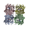 1zuxS S: Starting model for refinement |
|---|---|
| Similar structure data |


-Links
 PDBj
PDBj


- Assembly
| Deposited unit | 
| ||||||||
|---|---|---|---|---|---|---|---|---|---|
| 1 | 
| ||||||||
| 2 | 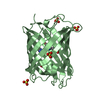
| ||||||||
| 3 | 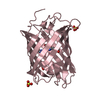
| ||||||||
| 4 | 
| ||||||||
| 5 |
| ||||||||
| Unit cell |
|
-Components
| #1: Protein | Mass: 26675.150 Da / Num. of mol.: 4 / Mutation: V123T T158H Source method: isolated from a genetically manipulated source Source: (gene. exp.) Lobophyllia hemprichii (invertebrata) / Plasmid: pQE32 / Production host:  Escherichia coli (E. coli) / References: UniProt: Q5S6Z9 Escherichia coli (E. coli) / References: UniProt: Q5S6Z9#2: Chemical | ChemComp-SO3 / Sulfite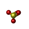  Mass: 80.063 Da / Num. of mol.: 4 / Source method: obtained synthetically / Formula: SO3 Mass: 80.063 Da / Num. of mol.: 4 / Source method: obtained synthetically / Formula: SO3#3: Chemical | ChemComp-SO4 / Sulfate  Mass: 96.063 Da / Num. of mol.: 15 / Source method: obtained synthetically / Formula: SO4 Mass: 96.063 Da / Num. of mol.: 15 / Source method: obtained synthetically / Formula: SO4#4: Water | ChemComp-HOH / | Water Mass: 18.015 Da / Num. of mol.: 382 / Source method: isolated from a natural source / Formula: H2O Mass: 18.015 Da / Num. of mol.: 382 / Source method: isolated from a natural source / Formula: H2OSequence details | HIS 62, TYR 63 AND GLY 64 CIRCULARIZ | |
|---|
-Experimental details
-Experiment
| Experiment | Method: X-RAY DIFFRACTION / Number of used crystals: 1 |
|---|
- Sample preparation
| Crystal | Density Matthews: 2.75 Å3/Da / Density % sol: 55.34 % |
|---|---|
| Crystal grow | Temperature: 298 K / Method: vapor diffusion, hanging drop / pH: 8.5 Details: 2.2 M ammonium sulfate, 0.1 M Tris-HCl, pH 8.5, VAPOR DIFFUSION, HANGING DROP, temperature 298.0K |
-Data collection
| Diffraction | Mean temperature: 100 K |
|---|---|
| Diffraction source | Source: SYNCHROTRON / Site: ESRF  / Beamline: ID14-3 / Wavelength: 0.931 Å / Beamline: ID14-3 / Wavelength: 0.931 Å |
| Detector | Type: ADSC QUANTUM 4 / Detector: CCD / Date: Jul 16, 2007 / Details: mirrors |
| Radiation | Monochromator: Si 111 CHANNEL / Protocol: SINGLE WAVELENGTH / Monochromatic (M) / Laue (L): M / Scattering type: x-ray |
| Radiation wavelength | Wavelength: 0.931 Å / Relative weight: 1 |
| Reflection | Resolution: 2.25→56.796 Å / Num. all: 80225 / Num. obs: 74471 / % possible obs: 92.8 % / Observed criterion σ(F): 2 / Observed criterion σ(I): 2 |
| Reflection shell | Resolution: 2.25→2.5 Å / % possible all: 99.5 |
- Processing
| Software |
| ||||||||||||||||||||||||||||||||||||||||||||||||||||||||||||||||||||||||||||||||||||||||||||||||||||||||||||||||||||||||||||||||||||||||||||||||||||||||||||||||||||||||||
|---|---|---|---|---|---|---|---|---|---|---|---|---|---|---|---|---|---|---|---|---|---|---|---|---|---|---|---|---|---|---|---|---|---|---|---|---|---|---|---|---|---|---|---|---|---|---|---|---|---|---|---|---|---|---|---|---|---|---|---|---|---|---|---|---|---|---|---|---|---|---|---|---|---|---|---|---|---|---|---|---|---|---|---|---|---|---|---|---|---|---|---|---|---|---|---|---|---|---|---|---|---|---|---|---|---|---|---|---|---|---|---|---|---|---|---|---|---|---|---|---|---|---|---|---|---|---|---|---|---|---|---|---|---|---|---|---|---|---|---|---|---|---|---|---|---|---|---|---|---|---|---|---|---|---|---|---|---|---|---|---|---|---|---|---|---|---|---|---|---|---|---|
| Refinement | Method to determine structure: MOLECULAR REPLACEMENT Starting model: PDB ENTRY 1ZUX Resolution: 2.25→48.41 Å / Cor.coef. Fo:Fc: 0.937 / Cor.coef. Fo:Fc free: 0.907 / SU B: 9.796 / SU ML: 0.228 / Cross valid method: THROUGHOUT / ESU R: 0.312 / ESU R Free: 0.247 / Stereochemistry target values: MAXIMUM LIKELIHOOD / Details: HYDROGENS HAVE BEEN ADDED IN THE RIDING POSITIONS
| ||||||||||||||||||||||||||||||||||||||||||||||||||||||||||||||||||||||||||||||||||||||||||||||||||||||||||||||||||||||||||||||||||||||||||||||||||||||||||||||||||||||||||
| Solvent computation | Ion probe radii: 0.8 Å / Shrinkage radii: 0.8 Å / VDW probe radii: 1.4 Å / Solvent model: MASK | ||||||||||||||||||||||||||||||||||||||||||||||||||||||||||||||||||||||||||||||||||||||||||||||||||||||||||||||||||||||||||||||||||||||||||||||||||||||||||||||||||||||||||
| Displacement parameters | Biso mean: 33.273 Å2
| ||||||||||||||||||||||||||||||||||||||||||||||||||||||||||||||||||||||||||||||||||||||||||||||||||||||||||||||||||||||||||||||||||||||||||||||||||||||||||||||||||||||||||
| Refinement step | Cycle: LAST / Resolution: 2.25→48.41 Å
| ||||||||||||||||||||||||||||||||||||||||||||||||||||||||||||||||||||||||||||||||||||||||||||||||||||||||||||||||||||||||||||||||||||||||||||||||||||||||||||||||||||||||||
| Refine LS restraints |
| ||||||||||||||||||||||||||||||||||||||||||||||||||||||||||||||||||||||||||||||||||||||||||||||||||||||||||||||||||||||||||||||||||||||||||||||||||||||||||||||||||||||||||
| LS refinement shell | Resolution: 2.25→2.308 Å / Total num. of bins used: 20
|
