 Movie
Movie Controller

[English] 日本語
 Yorodumi
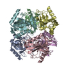
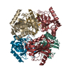
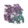
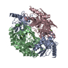
- PDB-3ld3: Crystal structure of inorganic phosphatase from anaplasma phagocy... -
+ Open data

- Basic information
| Entry | Database: PDB / ID: 3ld3 | ||||||
|---|---|---|---|---|---|---|---|
| Title | Crystal structure of inorganic phosphatase from anaplasma phagocytophilum at 1.75a resolution | ||||||
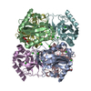
 Components Components | Inorganic pyrophosphatase | ||||||
 Keywords Keywords | HYDROLASE / Structural Genomics / Seattle Structural Genomics Center for Infectious Disease / SSGCID | ||||||
| Function / homology |  Function and homology informationinorganic diphosphatase / inorganic diphosphate phosphatase activity / phosphate-containing compound metabolic process / magnesium ion binding / cytoplasm Function and homology informationinorganic diphosphatase / inorganic diphosphate phosphatase activity / phosphate-containing compound metabolic process / magnesium ion binding / cytoplasmSimilarity search - Function | ||||||
| Biological species |  Anaplasma phagocytophilum (agent of human granulocytic ehrlichiosis) Anaplasma phagocytophilum (agent of human granulocytic ehrlichiosis) | ||||||
| Method | X-RAY DIFFRACTION / SYNCHROTRON / MOLECULAR REPLACEMENT / Resolution: 1.75 Å | ||||||
 Authors Authors | Seattle Structural Genomics Center for Infectious Disease (SSGCID) / Seattle Structural Genomics Center for Infectious Disease (SSGCID) | ||||||
 Citation Citation | Journal: To be Published Title: Crystal structure of inorganic phosphatase from anaplasma phagocytophilum at 1.75a resolution Authors: Seattle Structural Genomics Center for Infectious Disease (SSGCID) / Abendroth, J. / Davies, D. / Staker, B. | ||||||
| History |
|
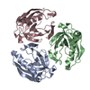
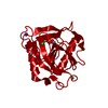
- Structure visualization
| Structure viewer | Molecule: MolmilJmol/JSmol |
|---|
- Downloads & links
-Download
| PDBx/mmCIF format | 3ld3.cif.gz | 85.5 KB | Display | PDBx/mmCIF format |
|---|---|---|---|---|
| PDB format | pdb3ld3.ent.gz | 64.5 KB | Display | PDB format |
| PDBx/mmJSON format | 3ld3.json.gz | Tree view | PDBx/mmJSON format | |
| Others |  Other downloads Other downloads |
-Validation report
| Arichive directory | https://data.pdbj.org/pub/pdb/validation_reports/ld/3ld3ftp://data.pdbj.org/pub/pdb/validation_reports/ld/3ld3 | HTTPS FTP |
|---|
-Related structure data
| Related structure data |  3fq3S S: Starting model for refinement |
|---|---|
| Similar structure data | |
| Other databases |

-Links
 PDBj
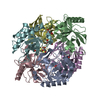
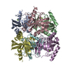
PDBj- Assembly
| Deposited unit | 
| ||||||||
|---|---|---|---|---|---|---|---|---|---|
| 1 | 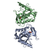
| ||||||||
| Unit cell |
|
-Components
| #1: Protein | Mass: 21886.742 Da / Num. of mol.: 2 Source method: isolated from a genetically manipulated source Source: (gene. exp.) Anaplasma phagocytophilum (agent of human granulocytic ehrlichiosis)Strain: HZ / Gene: ppa, APH_1101 / Plasmid: AVA0421 / Production host: Escherichia coli (E. coli) / Strain (production host): BL21(DE3) / References: UniProt: Q2GJ02, inorganic diphosphatase#2: Chemical | Phosphate  Mass: 94.971 Da / Num. of mol.: 2 / Source method: obtained synthetically / Formula: PO4 Mass: 94.971 Da / Num. of mol.: 2 / Source method: obtained synthetically / Formula: PO4#3: Water | ChemComp-HOH / | Water Mass: 18.015 Da / Num. of mol.: 256 / Source method: isolated from a natural source / Formula: H2O Mass: 18.015 Da / Num. of mol.: 256 / Source method: isolated from a natural source / Formula: H2O |
|---|
-Experimental details
-Experiment
| Experiment | Method: X-RAY DIFFRACTION / Number of used crystals: 1 |
|---|
- Sample preparation
| Crystal | Density Matthews: 2.36 Å3/Da / Density % sol: 47.92 % |
|---|---|
| Crystal grow | Temperature: 290 K / Method: vapor diffusion, sitting drop / pH: 4.2 Details: JCSG+ SCREEN CONDITION C6: 100MM PHOSPHATE/CITRATE PH 4.2, 40% PEG 300; ANPHA.01382.A AT 45MG/ ML, VAPOR DIFFUSION, SITTING DROP, TEMPERATURE 290K |
-Data collection
| Diffraction | Mean temperature: 100 K |
|---|---|
| Diffraction source | Source: SYNCHROTRON / Site: ALS  / Beamline: 5.0.3 / Wavelength: 0.9765 / Wavelength: 0.9765 Å / Beamline: 5.0.3 / Wavelength: 0.9765 / Wavelength: 0.9765 Å |
| Detector | Type: ADSC QUANTUM 315r / Detector: CCD / Date: Oct 26, 2009 / Details: Single crystal, cylindrically bent, Si(220) |
| Radiation | Protocol: SINGLE WAVELENGTH / Monochromatic (M) / Laue (L): M / Scattering type: x-ray |
| Radiation wavelength | Wavelength: 0.9765 Å / Relative weight: 1 |
| Reflection | Resolution: 1.75→20 Å / Num. all: 41592 / Num. obs: 41385 / % possible obs: 99.5 % / Observed criterion σ(F): 0 / Observed criterion σ(I): -3 / Redundancy: 10.5 % / Biso Wilson estimate: 30.82 Å2 / Rmerge(I) obs: 0.048 / Net I/σ(I): 30.24 |
| Reflection shell | Resolution: 1.75→1.8 Å / Redundancy: 8.8 % / Rmerge(I) obs: 0.529 / Mean I/σ(I) obs: 3.7 / Num. unique all: 3040 / % possible all: 99.9 |
- Processing
| Software |
| ||||||||||||||||||||||||||||||||||||||||||||||||||||||||||||||||||||||||||||||||||||||||||||||||||||||||||||||||||||||||||||||||||||||||||||||||||||||||||||||||||||||||||
|---|---|---|---|---|---|---|---|---|---|---|---|---|---|---|---|---|---|---|---|---|---|---|---|---|---|---|---|---|---|---|---|---|---|---|---|---|---|---|---|---|---|---|---|---|---|---|---|---|---|---|---|---|---|---|---|---|---|---|---|---|---|---|---|---|---|---|---|---|---|---|---|---|---|---|---|---|---|---|---|---|---|---|---|---|---|---|---|---|---|---|---|---|---|---|---|---|---|---|---|---|---|---|---|---|---|---|---|---|---|---|---|---|---|---|---|---|---|---|---|---|---|---|---|---|---|---|---|---|---|---|---|---|---|---|---|---|---|---|---|---|---|---|---|---|---|---|---|---|---|---|---|---|---|---|---|---|---|---|---|---|---|---|---|---|---|---|---|---|---|---|---|
| Refinement | Method to determine structure: MOLECULAR REPLACEMENT Starting model: PDB ENTRY 3FQ3 MODIFIED WITH CCP4 PROGRAM CHAINSAW Resolution: 1.75→19.96 Å / Cor.coef. Fo:Fc: 0.97 / Cor.coef. Fo:Fc free: 0.965 / SU B: 4.852 / SU ML: 0.068 / TLS residual ADP flag: LIKELY RESIDUAL / Isotropic thermal model: isotropic, TLS / Cross valid method: THROUGHOUT / σ(F): 0 / ESU R: 0.093 / ESU R Free: 0.089 / Stereochemistry target values: MAXIMUM LIKELIHOOD / Details: HYDROGENS HAVE BEEN ADDED IN THE RIDING POSITIONS
| ||||||||||||||||||||||||||||||||||||||||||||||||||||||||||||||||||||||||||||||||||||||||||||||||||||||||||||||||||||||||||||||||||||||||||||||||||||||||||||||||||||||||||
| Solvent computation | Ion probe radii: 0.8 Å / Shrinkage radii: 0.8 Å / VDW probe radii: 1.4 Å / Solvent model: BABINET MODEL WITH MASK | ||||||||||||||||||||||||||||||||||||||||||||||||||||||||||||||||||||||||||||||||||||||||||||||||||||||||||||||||||||||||||||||||||||||||||||||||||||||||||||||||||||||||||
| Displacement parameters | Biso mean: 12.61 Å2 | ||||||||||||||||||||||||||||||||||||||||||||||||||||||||||||||||||||||||||||||||||||||||||||||||||||||||||||||||||||||||||||||||||||||||||||||||||||||||||||||||||||||||||
| Refinement step | Cycle: LAST / Resolution: 1.75→19.96 Å
| ||||||||||||||||||||||||||||||||||||||||||||||||||||||||||||||||||||||||||||||||||||||||||||||||||||||||||||||||||||||||||||||||||||||||||||||||||||||||||||||||||||||||||
| Refine LS restraints |
| ||||||||||||||||||||||||||||||||||||||||||||||||||||||||||||||||||||||||||||||||||||||||||||||||||||||||||||||||||||||||||||||||||||||||||||||||||||||||||||||||||||||||||
| LS refinement shell | Resolution: 1.75→1.79 Å / Total num. of bins used: 20
| ||||||||||||||||||||||||||||||||||||||||||||||||||||||||||||||||||||||||||||||||||||||||||||||||||||||||||||||||||||||||||||||||||||||||||||||||||||||||||||||||||||||||||
| Refinement TLS params. | Method: refined / Refine-ID: X-RAY DIFFRACTION
| ||||||||||||||||||||||||||||||||||||||||||||||||||||||||||||||||||||||||||||||||||||||||||||||||||||||||||||||||||||||||||||||||||||||||||||||||||||||||||||||||||||||||||
| Refinement TLS group |
|
