 Movie
Movie Controller

[English] 日本語
 Yorodumi
- PDB-3d53: 2.2 A crystal structure of inorganic pyrophosphatase from Rickett... -
+ Open data

- Basic information
| Entry | Database: PDB / ID: 3d53 | ||||||
|---|---|---|---|---|---|---|---|
| Title | 2.2 A crystal structure of inorganic pyrophosphatase from Rickettsia prowazekii | ||||||
 Components Components | Inorganic pyrophosphatase | ||||||
 Keywords Keywords | HYDROLASE / RICKETTSIA / INORGANIC / PYROPHOSPHATASE / SEATTLE STRUCTURAL GENOMICS CENTER FOR INFECTIOUS DISEASE / SSGCID / Magnesium / Metal-binding | ||||||
| Function / homology |  Function and homology informationinorganic diphosphatase / inorganic diphosphate phosphatase activity / phosphate-containing compound metabolic process / magnesium ion binding / cytoplasm Function and homology informationinorganic diphosphatase / inorganic diphosphate phosphatase activity / phosphate-containing compound metabolic process / magnesium ion binding / cytoplasmSimilarity search - Function | ||||||
| Biological species |  Rickettsia prowazekii (bacteria) Rickettsia prowazekii (bacteria) | ||||||
| Method | X-RAY DIFFRACTION / SYNCHROTRON / MOLECULAR REPLACEMENT / molecular replacement / Resolution: 2.2 Å | ||||||
 Authors Authors | Seattle Structural Genomics Center for Infectious Disease (SSGCID) | ||||||
 Citation Citation | Journal: To be Published Title: 2.2 A crystal structure of inorganic pyrophosphatase from Rickettsia prowazekii. Authors: Seattle Structural Genomics Center for Infectious Disease (SSGCID) | ||||||
| History |
|
- Structure visualization
| Structure viewer | Molecule: MolmilJmol/JSmol |
|---|
- Downloads & links
-Download
| PDBx/mmCIF format | 3d53.cif.gz | 218.3 KB | Display | PDBx/mmCIF format |
|---|---|---|---|---|
| PDB format | pdb3d53.ent.gz | 175.1 KB | Display | PDB format |
| PDBx/mmJSON format | 3d53.json.gz | Tree view | PDBx/mmJSON format | |
| Others |  Other downloads Other downloads |
-Validation report
| Arichive directory | https://data.pdbj.org/pub/pdb/validation_reports/d5/3d53ftp://data.pdbj.org/pub/pdb/validation_reports/d5/3d53 | HTTPS FTP |
|---|
-Related structure data
| Related structure data |  2eipS S: Starting model for refinement |
|---|---|
| Similar structure data | |
| Other databases |


-Links
 PDBj
PDBj- Assembly
| Deposited unit | 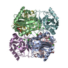
| ||||||||
|---|---|---|---|---|---|---|---|---|---|
| 1 |
| ||||||||
| Unit cell |
|
-Components
| #1: Protein | / Pyrophosphate phospho-hydrolase / PPase Mass: 19629.762 Da / Num. of mol.: 6 Source method: isolated from a genetically manipulated source Source: (gene. exp.) Rickettsia prowazekii (bacteria) / Strain: Madrid E / Gene: ppa, RP589 / Plasmid: pET28-SMT3 / Production host: Escherichia coli (E. coli) / Strain (production host): BL21(DE3) / References: UniProt: Q9ZCW5, inorganic diphosphatase#2: Chemical | ChemComp-CL / Chloride  Mass: 35.453 Da / Num. of mol.: 20 / Source method: obtained synthetically / Formula: Cl Mass: 35.453 Da / Num. of mol.: 20 / Source method: obtained synthetically / Formula: Cl#3: Chemical | Glycerol  Mass: 92.094 Da / Num. of mol.: 3 / Source method: obtained synthetically / Formula: C3H8O3 Mass: 92.094 Da / Num. of mol.: 3 / Source method: obtained synthetically / Formula: C3H8O3#4: Water | ChemComp-HOH / | Water Mass: 18.015 Da / Num. of mol.: 547 / Source method: isolated from a natural source / Formula: H2O Mass: 18.015 Da / Num. of mol.: 547 / Source method: isolated from a natural source / Formula: H2O |
|---|
-Experimental details
-Experiment
| Experiment | Method: X-RAY DIFFRACTION / Number of used crystals: 1 |
|---|
- Sample preparation
| Crystal | Density Matthews: 2.36 Å3/Da / Density % sol: 47.78 % |
|---|---|
| Crystal grow | Temperature: 293 K / Method: vapor diffusion / pH: 4.5 Details: 2.5M NaCl, 0.1M Na acetate pH 4.5, 0.2M Li2SO4, VAPOR DIFFUSION, temperature 293K |
-Data collection
| Diffraction | Mean temperature: 100 K | ||||||||||||||||||||||||||||||||||||||||||||||||||||||||||||||||||||||||||||||||||||||||
|---|---|---|---|---|---|---|---|---|---|---|---|---|---|---|---|---|---|---|---|---|---|---|---|---|---|---|---|---|---|---|---|---|---|---|---|---|---|---|---|---|---|---|---|---|---|---|---|---|---|---|---|---|---|---|---|---|---|---|---|---|---|---|---|---|---|---|---|---|---|---|---|---|---|---|---|---|---|---|---|---|---|---|---|---|---|---|---|---|---|
| Diffraction source | Source: SYNCHROTRON / Site: ALS  / Beamline: 5.0.1 / Wavelength: 0.977408 Å / Beamline: 5.0.1 / Wavelength: 0.977408 Å | ||||||||||||||||||||||||||||||||||||||||||||||||||||||||||||||||||||||||||||||||||||||||
| Detector | Type: ADSC QUANTUM 210 / Detector: CCD / Date: Apr 25, 2008 / Details: Adjustable focusing mirrors | ||||||||||||||||||||||||||||||||||||||||||||||||||||||||||||||||||||||||||||||||||||||||
| Radiation | Monochromator: Single crystal, cylindrically bent, Si(220) / Protocol: SINGLE WAVELENGTH / Monochromatic (M) / Laue (L): M / Scattering type: x-ray | ||||||||||||||||||||||||||||||||||||||||||||||||||||||||||||||||||||||||||||||||||||||||
| Radiation wavelength | Wavelength: 0.977408 Å / Relative weight: 1 | ||||||||||||||||||||||||||||||||||||||||||||||||||||||||||||||||||||||||||||||||||||||||
| Reflection | Resolution: 2.2→34.94 Å / Num. obs: 56518 / % possible obs: 98.8 % / Redundancy: 6.2 % / Rmerge(I) obs: 0.132 / Rsym value: 0.132 / Net I/σ(I): 5 | ||||||||||||||||||||||||||||||||||||||||||||||||||||||||||||||||||||||||||||||||||||||||
| Reflection shell | Diffraction-ID: 1
|
-Phasing
| Phasing | Method: molecular replacement | |||||||||
|---|---|---|---|---|---|---|---|---|---|---|
| Phasing MR | Model details: Phaser MODE: MR_AUTO
|
- Processing
| Software |
| ||||||||||||||||||||||||||||||||||||||||||||||||||||||||||||||||||||||||||||||||||||||||||
|---|---|---|---|---|---|---|---|---|---|---|---|---|---|---|---|---|---|---|---|---|---|---|---|---|---|---|---|---|---|---|---|---|---|---|---|---|---|---|---|---|---|---|---|---|---|---|---|---|---|---|---|---|---|---|---|---|---|---|---|---|---|---|---|---|---|---|---|---|---|---|---|---|---|---|---|---|---|---|---|---|---|---|---|---|---|---|---|---|---|---|---|
| Refinement | Method to determine structure: MOLECULAR REPLACEMENT Starting model: PDB entry 2EIP Resolution: 2.2→33.56 Å / Cor.coef. Fo:Fc: 0.943 / Cor.coef. Fo:Fc free: 0.901 / SU B: 7.013 / SU ML: 0.18 / Cross valid method: THROUGHOUT / σ(F): 0 / ESU R: 0.332 / ESU R Free: 0.254 / Stereochemistry target values: MAXIMUM LIKELIHOOD
| ||||||||||||||||||||||||||||||||||||||||||||||||||||||||||||||||||||||||||||||||||||||||||
| Solvent computation | Ion probe radii: 0.8 Å / Shrinkage radii: 0.8 Å / VDW probe radii: 1.2 Å / Solvent model: MASK | ||||||||||||||||||||||||||||||||||||||||||||||||||||||||||||||||||||||||||||||||||||||||||
| Displacement parameters | Biso mean: 20.576 Å2
| ||||||||||||||||||||||||||||||||||||||||||||||||||||||||||||||||||||||||||||||||||||||||||
| Refinement step | Cycle: LAST / Resolution: 2.2→33.56 Å
| ||||||||||||||||||||||||||||||||||||||||||||||||||||||||||||||||||||||||||||||||||||||||||
| Refine LS restraints |
| ||||||||||||||||||||||||||||||||||||||||||||||||||||||||||||||||||||||||||||||||||||||||||
| LS refinement shell | Resolution: 2.2→2.257 Å / Total num. of bins used: 20
|
