 Movie
Movie Controller

[English] 日本語
 Yorodumi
- PDB-1ude: Crystal structure of the Inorganic pyrophosphatase from the hyper... -
+ Open data

- Basic information
| Entry | Database: PDB / ID: 1ude | ||||||
|---|---|---|---|---|---|---|---|
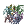
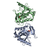
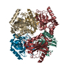
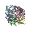
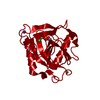
| Title | Crystal structure of the Inorganic pyrophosphatase from the hyperthermophilic archaeon Pyrococcus horikoshii OT3 | ||||||
 Components Components | Inorganic pyrophosphatase | ||||||
 Keywords Keywords | HYDROLASE / inorganic pyrophosphatase X-ray crystallographic analysis | ||||||
| Function / homology |  Function and homology informationinorganic diphosphatase / inorganic diphosphate phosphatase activity / phosphate-containing compound metabolic process / magnesium ion binding / cytoplasm Function and homology informationinorganic diphosphatase / inorganic diphosphate phosphatase activity / phosphate-containing compound metabolic process / magnesium ion binding / cytoplasmSimilarity search - Function | ||||||
| Biological species |   Pyrococcus horikoshii (archaea) Pyrococcus horikoshii (archaea) | ||||||
| Method | X-RAY DIFFRACTION / MOLECULAR REPLACEMENT / Resolution: 2.66 Å | ||||||
 Authors Authors | Liu, B. / Gao, R. / Zhou, W. / Bartlam, M. / Rao, Z. | ||||||
 Citation Citation | Journal: Biophys.J. / Year: 2004 Title: Crystal structure of the hyperthermophilic inorganic pyrophosphatase from the archaeon Pyrococcus horikoshii. Authors: Liu, B. / Bartlam, M. / Gao, R. / Zhou, W. / Pang, H. / Liu, Y. / Feng, Y. / Rao, Z. | ||||||
| History |
|
- Structure visualization
| Structure viewer | Molecule: MolmilJmol/JSmol |
|---|
- Downloads & links
-Download
| PDBx/mmCIF format | 1ude.cif.gz | 114.7 KB | Display | PDBx/mmCIF format |
|---|---|---|---|---|
| PDB format | pdb1ude.ent.gz | 90 KB | Display | PDB format |
| PDBx/mmJSON format | 1ude.json.gz | Tree view | PDBx/mmJSON format | |
| Others |  Other downloads Other downloads |
-Validation report
| Arichive directory | https://data.pdbj.org/pub/pdb/validation_reports/ud/1udeftp://data.pdbj.org/pub/pdb/validation_reports/ud/1ude | HTTPS FTP |
|---|
-Related structure data
| Similar structure data |
|---|


-Links
 PDBj
PDBj- Assembly
| Deposited unit | 
| ||||||||
|---|---|---|---|---|---|---|---|---|---|
| 1 | 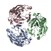
| ||||||||
| Unit cell |
| ||||||||
| Details | The biological assembly is a hexamer generated from the trimer in the asymmetric unit by crystallographic quadratic operations |
-Components
| #1: Protein | Mass: 22803.168 Da / Num. of mol.: 3 Source method: isolated from a genetically manipulated source Source: (gene. exp.) Pyrococcus horikoshii (archaea) / Gene: PPA / Plasmid: PET15b / Production host:  Escherichia coli (E. coli) / Strain (production host): BE21 / References: UniProt: O59570, inorganic diphosphatase Escherichia coli (E. coli) / Strain (production host): BE21 / References: UniProt: O59570, inorganic diphosphatase#2: Water | ChemComp-HOH / | Water Mass: 18.015 Da / Num. of mol.: 93 / Source method: isolated from a natural source / Formula: H2O Mass: 18.015 Da / Num. of mol.: 93 / Source method: isolated from a natural source / Formula: H2O |
|---|
-Experimental details
-Experiment
| Experiment | Method: X-RAY DIFFRACTION / Number of used crystals: 1 |
|---|
- Sample preparation
| Crystal | Density Matthews: 2.44 Å3/Da / Density % sol: 49.1 % | |||||||||||||||||||||||||||||||||||
|---|---|---|---|---|---|---|---|---|---|---|---|---|---|---|---|---|---|---|---|---|---|---|---|---|---|---|---|---|---|---|---|---|---|---|---|---|
| Crystal grow | Temperature: 291 K / Method: vapor diffusion, hanging drop / pH: 5 Details: PEG 4000, Na Acetete, pH 5.0, VAPOR DIFFUSION, HANGING DROP, temperature 291K | |||||||||||||||||||||||||||||||||||
| Crystal grow | *PLUS Method: vapor diffusion, hanging drop / PH range low: 5.2 / PH range high: 5 | |||||||||||||||||||||||||||||||||||
| Components of the solutions | *PLUS
|
-Data collection
| Diffraction | Mean temperature: 100 K |
|---|---|
| Diffraction source | Source: ROTATING ANODE / Type: RIGAKU / Wavelength: 1.5418 Å |
| Detector | Type: MARRESEARCH / Detector: IMAGE PLATE / Date: Jun 1, 2002 |
| Radiation | Protocol: SINGLE WAVELENGTH / Monochromatic (M) / Laue (L): M / Scattering type: x-ray |
| Radiation wavelength | Wavelength: 1.5418 Å / Relative weight: 1 |
| Reflection | Resolution: 2.66→50 Å / Num. obs: 17030 / % possible obs: 99.8 % / Observed criterion σ(F): 2 / Observed criterion σ(I): 2 / Redundancy: 6.93 % / Rmerge(I) obs: 0.075 |
| Reflection shell | Resolution: 2.66→2.9 Å / % possible all: 98 |
| Reflection | *PLUS Num. measured all: 118041 |
| Reflection shell | *PLUS % possible obs: 98 % / Rmerge(I) obs: 0.139 / Mean I/σ(I) obs: 4.3 |
- Processing
| Software |
| |||||||||||||||||||||||||
|---|---|---|---|---|---|---|---|---|---|---|---|---|---|---|---|---|---|---|---|---|---|---|---|---|---|---|
| Refinement | Method to determine structure: MOLECULAR REPLACEMENT / Resolution: 2.66→50 Å / Rfactor Rfree error: 0.006 / Cross valid method: THROUGHOUT / σ(F): 0 / Stereochemistry target values: Engh & Huber Details: inorganic pyrophosphatase Sulfolobus acidocaldarius (S-PPase)
| |||||||||||||||||||||||||
| Refinement step | Cycle: LAST / Resolution: 2.66→50 Å
| |||||||||||||||||||||||||
| Refine LS restraints |
| |||||||||||||||||||||||||
| LS refinement shell | Resolution: 2.66→2.83 Å / Rfactor Rfree error: 0.018 / Total num. of bins used: 6
| |||||||||||||||||||||||||
| Refinement | *PLUS Num. reflection obs: 117986 | |||||||||||||||||||||||||
| Solvent computation | *PLUS | |||||||||||||||||||||||||
| Displacement parameters | *PLUS | |||||||||||||||||||||||||
| Refine LS restraints | *PLUS
|
