 Movie
Movie Controller


+ Open data

- Basic information
| Entry | Database: PDB / ID: 3l57 | ||||||
|---|---|---|---|---|---|---|---|
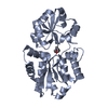
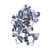
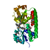
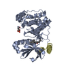
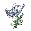
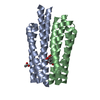
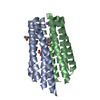
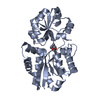
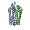
| Title | Crystal Structure of the Plasmid pCU1 TraI Relaxase Domain | ||||||
 Components Components | Mobilization protein TraI | ||||||
 Keywords Keywords | HYDROLASE / TrwC superfamily of relaxase enzymes / conjugative relaxase / Mob class relaxase / conjugal nickase / histidine triad / HUH+H motif | ||||||
| Function / homology |  Function and homology information Function and homology information | ||||||
| Biological species |  Escherichia coli (E. coli) Escherichia coli (E. coli) | ||||||
| Method | X-RAY DIFFRACTION / SYNCHROTRON / MOLECULAR REPLACEMENT / molecular replacement / Resolution: 2.293 Å | ||||||
 Authors Authors | Redinbo, M.R. / Nash, R.P. | ||||||
 Citation Citation | Journal: Nucleic Acids Res. / Year: 2010 Title: The mechanism and control of DNA transfer by the conjugative relaxase of resistance plasmid pCU1. Authors: Nash, R.P. / Habibi, S. / Cheng, Y. / Lujan, S.A. / Redinbo, M.R. | ||||||
| History |
|
- Structure visualization
| Structure viewer | Molecule: MolmilJmol/JSmol |
|---|
- Downloads & links
-Download
| PDBx/mmCIF format | 3l57.cif.gz | 199.9 KB | Display | PDBx/mmCIF format |
|---|---|---|---|---|
| PDB format | pdb3l57.ent.gz | 158.8 KB | Display | PDB format |
| PDBx/mmJSON format | 3l57.json.gz | Tree view | PDBx/mmJSON format | |
| Others |  Other downloads Other downloads |
-Validation report
| Arichive directory | https://data.pdbj.org/pub/pdb/validation_reports/l5/3l57ftp://data.pdbj.org/pub/pdb/validation_reports/l5/3l57 | HTTPS FTP |
|---|
-Related structure data
| Related structure data |  3l6tC  1omhS S: Starting model for refinement C: citing same article ( |
|---|---|
| Similar structure data |

-Links
 PDBj
PDBj

- Assembly
| Deposited unit | 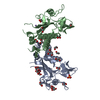
| ||||||||
|---|---|---|---|---|---|---|---|---|---|
| 1 | 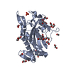
| ||||||||
| 2 | 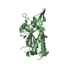
| ||||||||
| Unit cell |
|
-Components
-Protein , 1 types, 2 molecules AB
| #1: Protein | / TraI Mass: 33951.863 Da / Num. of mol.: 2 / Fragment: Relaxase Domain Source method: isolated from a genetically manipulated source Source: (gene. exp.) Escherichia coli (E. coli) / Gene: pMUR050_047, traI / Plasmid: pTYB2 / Production host: Escherichia coli (E. coli) / Strain (production host): BL21(DE3)References: UniProt: Q9X4G2, Hydrolases; Acting on acid anhydrides; In phosphorus-containing anhydrides |
|---|
-Non-polymers , 5 types, 249 molecules 


| #2: Chemical | Manganese Mass: 54.938 Da / Num. of mol.: 2 / Source method: obtained synthetically / Formula: Mn Mass: 54.938 Da / Num. of mol.: 2 / Source method: obtained synthetically / Formula: Mn#3: Chemical | ChemComp-EDO / Ethylene glycol Mass: 62.068 Da / Num. of mol.: 26 / Source method: obtained synthetically / Formula: C2H6O2 Mass: 62.068 Da / Num. of mol.: 26 / Source method: obtained synthetically / Formula: C2H6O2#4: Chemical | ChemComp-PO4 / | Phosphate Mass: 94.971 Da / Num. of mol.: 1 / Source method: obtained synthetically / Formula: PO4 Mass: 94.971 Da / Num. of mol.: 1 / Source method: obtained synthetically / Formula: PO4#5: Chemical | ChemComp-CIT / | Citric acid Mass: 192.124 Da / Num. of mol.: 1 / Source method: obtained synthetically / Formula: C6H8O7 Mass: 192.124 Da / Num. of mol.: 1 / Source method: obtained synthetically / Formula: C6H8O7#6: Water | ChemComp-HOH / | WaterMass: 18.015 Da / Num. of mol.: 219 / Source method: isolated from a natural source / Formula: H2O |
|---|
-Experimental details
-Experiment
| Experiment | Method: X-RAY DIFFRACTION / Number of used crystals: 1 |
|---|
- Sample preparation
| Crystal | Density Matthews: 1.88 Å3/Da / Density % sol: 34.46 % |
|---|---|
| Crystal grow | Temperature: 293.15 K / Method: vapor diffusion, hanging drop Details: 250mM triNa citrate, 22% PEG 3350, 5mM MgCl2, vapor diffusion, hanging drop, temperature 293.15K |
-Data collection
| Diffraction | Mean temperature: 100 K |
|---|---|
| Diffraction source | Source: SYNCHROTRON / Site: APS  / Beamline: 22-ID / Wavelength: 0.999 Å / Beamline: 22-ID / Wavelength: 0.999 Å |
| Detector | Type: MAR scanner 300 mm plate / Detector: IMAGE PLATE / Date: Feb 1, 2008 / Details: Rosenbaum-Rock vertical focusing mirror |
| Radiation | Monochromator: Rosenbaum-Rock monochromator high-resolution double-crystal Si(220) Protocol: SINGLE WAVELENGTH / Monochromatic (M) / Laue (L): M / Scattering type: x-ray |
| Radiation wavelength | Wavelength: 0.999 Å / Relative weight: 1 |
| Reflection | Resolution: 2.29→49.999 Å / Num. all: 22781 / Num. obs: 22081 / % possible obs: 96.6 % / Observed criterion σ(I): -3 / Biso Wilson estimate: 45.743 Å2 / Rsym value: 0.054 / Net I/σ(I): 18.25 |
| Reflection shell | Resolution: 2.29→2.43 Å / Mean I/σ(I) obs: 4.5 / Num. measured obs: 12219 / Num. unique all: 3362 / Num. unique obs: 3362 / Rsym value: 0.401 / % possible all: 91.8 |
-Phasing
| Phasing | Method: molecular replacement | |||||||||
|---|---|---|---|---|---|---|---|---|---|---|
| Phasing MR | Model details: Phaser MODE: MR_AUTO
|
- Processing
| Software |
| ||||||||||||||||||||||||||||||||||||||||||||||||||||||||||||||||||||||||
|---|---|---|---|---|---|---|---|---|---|---|---|---|---|---|---|---|---|---|---|---|---|---|---|---|---|---|---|---|---|---|---|---|---|---|---|---|---|---|---|---|---|---|---|---|---|---|---|---|---|---|---|---|---|---|---|---|---|---|---|---|---|---|---|---|---|---|---|---|---|---|---|---|---|
| Refinement | Method to determine structure: MOLECULAR REPLACEMENT Starting model: PDB entry 1omh, Plasmid R388 TrwC Relaxase Domain Resolution: 2.293→48.492 Å / Occupancy max: 1 / Occupancy min: 0.2 / FOM work R set: 0.842 / SU ML: 0.08 / Isotropic thermal model: isotropic + TLS / Cross valid method: THROUGHOUT / σ(F): 1.99 / Stereochemistry target values: ML
| ||||||||||||||||||||||||||||||||||||||||||||||||||||||||||||||||||||||||
| Solvent computation | Shrinkage radii: 0.9 Å / VDW probe radii: 1.11 Å / Solvent model: FLAT BULK SOLVENT MODEL / Bsol: 65.392 Å2 / ksol: 0.338 e/Å3 | ||||||||||||||||||||||||||||||||||||||||||||||||||||||||||||||||||||||||
| Displacement parameters | Biso max: 107.69 Å2 / Biso mean: 47.255 Å2 / Biso min: 20.74 Å2
| ||||||||||||||||||||||||||||||||||||||||||||||||||||||||||||||||||||||||
| Refinement step | Cycle: LAST / Resolution: 2.293→48.492 Å
| ||||||||||||||||||||||||||||||||||||||||||||||||||||||||||||||||||||||||
| Refine LS restraints |
| ||||||||||||||||||||||||||||||||||||||||||||||||||||||||||||||||||||||||
| LS refinement shell | Refine-ID: X-RAY DIFFRACTION / Total num. of bins used: 8
| ||||||||||||||||||||||||||||||||||||||||||||||||||||||||||||||||||||||||
| Refinement TLS params. | Method: refined / Origin x: 36.0064 Å / Origin y: 32.0627 Å / Origin z: 25.829 Å
| ||||||||||||||||||||||||||||||||||||||||||||||||||||||||||||||||||||||||
| Refinement TLS group |
|
