 Movie
Movie Controller


+ Open data

- Basic information
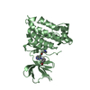
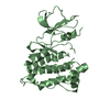
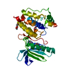
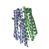
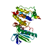
| Entry | Database: PDB / ID: 3kf6 | ||||||
|---|---|---|---|---|---|---|---|
| Title | Crystal structure of S. pombe Stn1-ten1 complex | ||||||
 Components Components |
| ||||||
 Keywords Keywords |  STRUCTURAL PROTEIN / OB fold / Chromosomal protein / DNA-binding / Nucleus / Telomere STRUCTURAL PROTEIN / OB fold / Chromosomal protein / DNA-binding / Nucleus / Telomere | ||||||
| Function / homology |  Function and homology informationCST complex / telomere cap complex / chromosome, telomeric repeat region / single-stranded telomeric DNA binding / telomere capping / negative regulation of telomere maintenance via telomerase / nucleus Function and homology informationCST complex / telomere cap complex / chromosome, telomeric repeat region / single-stranded telomeric DNA binding / telomere capping / negative regulation of telomere maintenance via telomerase / nucleusSimilarity search - Function | ||||||
| Biological species |  Schizosaccharomyces pombe (fission yeast) Schizosaccharomyces pombe (fission yeast) | ||||||
| Method | X-RAY DIFFRACTION / SYNCHROTRON / SAD / Resolution: 1.65 Å | ||||||
 Authors Authors | Sun, J. / Yu, E.Y. / Yang, Y.T. / Confer, L.A. / Sun, S.H. / Wan, K. / Lue, N.F. / Lei, M. | ||||||
 Citation Citation | Journal: Genes Dev. / Year: 2009 Title: Stn1-Ten1 is an Rpa2-Rpa3-like complex at telomeres. Authors: Sun, J. / Yu, E.Y. / Yang, Y. / Confer, L.A. / Sun, S.H. / Wan, K. / Lue, N.F. / Lei, M. | ||||||
| History |
|
- Structure visualization
| Structure viewer | Molecule: MolmilJmol/JSmol |
|---|
- Downloads & links
-Download
| PDBx/mmCIF format | 3kf6.cif.gz | 64.8 KB | Display | PDBx/mmCIF format |
|---|---|---|---|---|
| PDB format | pdb3kf6.ent.gz | 47.6 KB | Display | PDB format |
| PDBx/mmJSON format | 3kf6.json.gz | Tree view | PDBx/mmJSON format | |
| Others |  Other downloads Other downloads |
-Validation report
| Arichive directory | https://data.pdbj.org/pub/pdb/validation_reports/kf/3kf6ftp://data.pdbj.org/pub/pdb/validation_reports/kf/3kf6 | HTTPS FTP |
|---|
-Related structure data


-Links
 PDBj
PDBj
- Assembly
| Deposited unit | 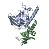
| ||||||||
|---|---|---|---|---|---|---|---|---|---|
| 1 |
| ||||||||
| Unit cell |
| ||||||||
| Components on special symmetry positions |
|
-Components
| #1: Protein | Mass: 18755.656 Da / Num. of mol.: 1 / Fragment: N-terminal fragment: UNP residues 2-159 Source method: isolated from a genetically manipulated source Source: (gene. exp.) Schizosaccharomyces pombe (fission yeast)Gene: stn1, SPBC409.12c / Plasmid: pET28b / Production host:  Escherichia coli (E. coli) / Strain (production host): BL21 / References: UniProt: Q0E7J7 Escherichia coli (E. coli) / Strain (production host): BL21 / References: UniProt: Q0E7J7 |
|---|---|
| #2: Protein | Mass: 11772.649 Da / Num. of mol.: 1 / Fragment: UNP residues 2-102 Source method: isolated from a genetically manipulated source Source: (gene. exp.) Schizosaccharomyces pombe (fission yeast)Gene: ten1, SPCC1393.14 / Plasmid: pET28b / Production host: Escherichia coli (E. coli) / Strain (production host): BL21 / References: UniProt: P0C5Y7 |
| #3: Water | ChemComp-HOH / Water Mass: 18.015 Da / Num. of mol.: 191 / Source method: isolated from a natural source / Formula: H2O Mass: 18.015 Da / Num. of mol.: 191 / Source method: isolated from a natural source / Formula: H2O |
-Experimental details
-Experiment
| Experiment | Method: X-RAY DIFFRACTION |
|---|
- Sample preparation
| Crystal | Density Matthews: 2.03 Å3/Da / Density % sol: 39.42 % |
|---|---|
| Crystal grow | Temperature: 277 K / Method: vapor diffusion, sitting drop / pH: 5.6 Details: 12% PEG 4000, 12% Isopropanol, 0.1 M Sodium citrate pH 5.6, 5 mM DTT, VAPOR DIFFUSION, SITTING DROP, temperature 277K |
-Data collection
| Diffraction | Mean temperature: 100 K |
|---|---|
| Diffraction source | Source: SYNCHROTRON / Site: APS  / Beamline: 21-ID-D / Wavelength: 1 Å / Beamline: 21-ID-D / Wavelength: 1 Å |
| Detector | Type: MARMOSAIC 225 mm CCD / Detector: CCD / Date: Jun 5, 2008 |
| Radiation | Protocol: SINGLE WAVELENGTH / Monochromatic (M) / Laue (L): M / Scattering type: x-ray |
| Radiation wavelength | Wavelength: 1 Å / Relative weight: 1 |
| Reflection | Resolution: 1.51→50 Å / Num. all: 35289 / Num. obs: 30813 / % possible obs: 88 % / Redundancy: 12.2 % / Rmerge(I) obs: 0.075 / Net I/σ(I): 42.3 |
- Processing
| Software |
| ||||||||||||||||||||
|---|---|---|---|---|---|---|---|---|---|---|---|---|---|---|---|---|---|---|---|---|---|
| Refinement | Method to determine structure: SAD / Resolution: 1.65→50 Å
| ||||||||||||||||||||
| Refinement step | Cycle: LAST / Resolution: 1.65→50 Å
|
