 Movie
Movie Controller

[English] 日本語
 Yorodumi
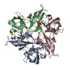
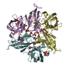
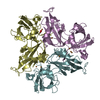
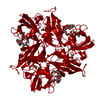
- PDB-3kw8: Two-domain laccase from Streptomyces coelicolor at 2.3 A resolution -
+ Open data

- Basic information
| Entry | Database: PDB / ID: 3kw8 | ||||||
|---|---|---|---|---|---|---|---|
| Title | Two-domain laccase from Streptomyces coelicolor at 2.3 A resolution | ||||||
 Components Components | Putative copper oxidase | ||||||
 Keywords Keywords |  OXIDOREDUCTASE / two-domain laccase / multicopper blue protein OXIDOREDUCTASE / two-domain laccase / multicopper blue protein | ||||||
| Function / homology |  Function and homology information Function and homology information | ||||||
| Biological species |  Streptomyces coelicolor (bacteria) Streptomyces coelicolor (bacteria) | ||||||
| Method | X-RAY DIFFRACTION / SYNCHROTRON / MOLECULAR REPLACEMENT / Resolution: 2.29 Å | ||||||
 Authors Authors | Skalova, T. / Dohnalek, J. / Kolenko, P. / Duskova, J. / Stepankova, A. / Hasek, J. / Ostergaard, L.H. / Ostergaard, P.R. | ||||||
 Citation Citation | Journal: Acta Crystallogr.,Sect.F / Year: 2011 Title: Structure of laccase from Streptomyces coelicolor after soaking with potassium hexacyanoferrate and at an improved resolution of 2.3 A Authors: Skalova, T. / Duskova, J. / Hasek, J. / Stepankova, A. / Koval, T. / Ostergaard, L.H. / Dohnalek, J. #1: Journal: J.Mol.Biol. / Year: 2009Title: The structure of the small laccase from Streptomyces coelicolor reveals a link between laccases and nitrite reductases Authors: Skalova, T. / Dohnalek, J. / Ostergaard, L.H. / Ostergaard, P.R. / Kolenko, P. / Duskova, J. / Stepankova, A. / Hasek, J. #2: Journal: Acta Crystallogr.,Sect.F / Year: 2007 Title: Crystallization and preliminary X-ray diffraction analysis of the small laccase from Streptomyces coelicolor Authors: Skalova, T. / Dohnalek, J. / Ostergaard, L.H. / Ostergaard, P.R. / Kolenko, P. / Duskova, J. / Hasek, J. | ||||||
| History |
|
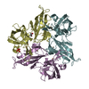
- Structure visualization
| Structure viewer | Molecule: MolmilJmol/JSmol |
|---|
- Downloads & links
-Download
| PDBx/mmCIF format | 3kw8.cif.gz | 77.3 KB | Display | PDBx/mmCIF format |
|---|---|---|---|---|
| PDB format | pdb3kw8.ent.gz | 55.8 KB | Display | PDB format |
| PDBx/mmJSON format | 3kw8.json.gz | Tree view | PDBx/mmJSON format | |
| Others |  Other downloads Other downloads |
-Validation report
| Arichive directory | https://data.pdbj.org/pub/pdb/validation_reports/kw/3kw8ftp://data.pdbj.org/pub/pdb/validation_reports/kw/3kw8 | HTTPS FTP |
|---|
-Related structure data
| Related structure data |  3cg8S S: Starting model for refinement |
|---|---|
| Similar structure data |


-Links
 PDBj
PDBj

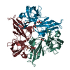
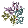
- Assembly
| Deposited unit | 
| ||||||||||||
|---|---|---|---|---|---|---|---|---|---|---|---|---|---|
| 1 | 
| ||||||||||||
| Unit cell |
| ||||||||||||
| Components on special symmetry positions |
|
-Components
-Protein , 1 types, 1 molecules A
| #1: Protein | Mass: 30378.855 Da / Num. of mol.: 1 / Fragment: residues 42-317 Source method: isolated from a genetically manipulated source Details: TAKA amylase promoter / Source: (gene. exp.) Streptomyces coelicolor (bacteria) / Strain: A3(2) / Gene: SC4C6.22 / Plasmid: PLAQ029 / Production host:  Aspergillus oryzae (mold) / Strain (production host): TOC1512 / References: UniProt: Q9XAL8, laccase Aspergillus oryzae (mold) / Strain (production host): TOC1512 / References: UniProt: Q9XAL8, laccase |
|---|
-Non-polymers , 6 types, 246 molecules 


| #2: Chemical | ChemComp-CU / Copper Mass: 63.546 Da / Num. of mol.: 4 / Source method: obtained synthetically / Formula: Cu Mass: 63.546 Da / Num. of mol.: 4 / Source method: obtained synthetically / Formula: Cu#3: Chemical | ChemComp-FE / | Iron Mass: 55.845 Da / Num. of mol.: 1 / Source method: obtained synthetically / Formula: Fe Mass: 55.845 Da / Num. of mol.: 1 / Source method: obtained synthetically / Formula: Fe#4: Chemical |  Mass: 22.990 Da / Num. of mol.: 2 / Source method: obtained synthetically / Formula: Na Mass: 22.990 Da / Num. of mol.: 2 / Source method: obtained synthetically / Formula: Na#5: Chemical | Polyethylene glycol Mass: 194.226 Da / Num. of mol.: 2 / Source method: obtained synthetically / Formula: C8H18O5 / Comment: precipitant*YM Mass: 194.226 Da / Num. of mol.: 2 / Source method: obtained synthetically / Formula: C8H18O5 / Comment: precipitant*YM#6: Chemical | Polyethylene glycol Mass: 150.173 Da / Num. of mol.: 3 / Source method: obtained synthetically / Formula: C6H14O4 Mass: 150.173 Da / Num. of mol.: 3 / Source method: obtained synthetically / Formula: C6H14O4#7: Water | ChemComp-HOH / | WaterMass: 18.015 Da / Num. of mol.: 234 / Source method: isolated from a natural source / Formula: H2O |
|---|
-Details
| Nonpolymer details | FE IN THIS STRUCTURE IS A PART OF A HEXACYANOF |
|---|
-Experimental details
-Experiment
| Experiment | Method: X-RAY DIFFRACTION / Number of used crystals: 1 |
|---|
- Sample preparation
| Crystal | Density Matthews: 7.569955 Å3/Da / Density % sol: 83.751556 % |
|---|---|
| Crystal grow | Temperature: 298 K / Method: vapor diffusion, hanging drop / pH: 9 Details: 40 %(v/v) PEG 550 monomethyl ether, 0.1M NaCl, 0.1M glycine, 50mM potassium hexacyanoferrate , pH 9.0, VAPOR DIFFUSION, HANGING DROP, temperature 298K |
-Data collection
| Diffraction | Mean temperature: 100 K |
|---|---|
| Diffraction source | Source: SYNCHROTRON / Site: BESSY  / Beamline: 14.1 / Wavelength: 0.91841 Å / Beamline: 14.1 / Wavelength: 0.91841 Å |
| Detector | Type: MARMOSAIC 225 mm CCD / Detector: CCD / Date: Apr 18, 2007 / Details: mirrors |
| Radiation | Monochromator: Si-111 crystal / Protocol: SINGLE WAVELENGTH / Monochromatic (M) / Laue (L): M / Scattering type: x-ray |
| Radiation wavelength | Wavelength: 0.91841 Å / Relative weight: 1 |
| Reflection | Resolution: 2.29→50 Å / Num. all: 42751 / Num. obs: 42751 / % possible obs: 100 % / Redundancy: 24.1 % / Biso Wilson estimate: 43.6 Å2 / Rmerge(I) obs: 0.114 / Net I/σ(I): 33.9 |
| Reflection shell | Resolution: 2.3→2.34 Å / Redundancy: 24.6 % / Rmerge(I) obs: 0.605 / Mean I/σ(I) obs: 6.3 / Num. unique all: 2077 / % possible all: 100 |
- Processing
| Software |
| ||||||||||||||||||||||||||||||||||||||||||||||||||||||||||||||||||||||||||||||||||||||||||||||||||||||||||||||||||||||||||||||||||||||||||||||||||||||||||||||||||||||||||
|---|---|---|---|---|---|---|---|---|---|---|---|---|---|---|---|---|---|---|---|---|---|---|---|---|---|---|---|---|---|---|---|---|---|---|---|---|---|---|---|---|---|---|---|---|---|---|---|---|---|---|---|---|---|---|---|---|---|---|---|---|---|---|---|---|---|---|---|---|---|---|---|---|---|---|---|---|---|---|---|---|---|---|---|---|---|---|---|---|---|---|---|---|---|---|---|---|---|---|---|---|---|---|---|---|---|---|---|---|---|---|---|---|---|---|---|---|---|---|---|---|---|---|---|---|---|---|---|---|---|---|---|---|---|---|---|---|---|---|---|---|---|---|---|---|---|---|---|---|---|---|---|---|---|---|---|---|---|---|---|---|---|---|---|---|---|---|---|---|---|---|---|
| Refinement | Method to determine structure: MOLECULAR REPLACEMENT Starting model: PDB ENTRY 3CG8 Resolution: 2.29→38.56 Å / Cor.coef. Fo:Fc: 0.963 / Cor.coef. Fo:Fc free: 0.953 / SU B: 2.041 / SU ML: 0.052 / Cross valid method: THROUGHOUT / ESU R: 0.104 / ESU R Free: 0.101 / Stereochemistry target values: MAXIMUM LIKELIHOOD / Details: HYDROGENS WERE REFINED IN THE RIDING POSITIONS
| ||||||||||||||||||||||||||||||||||||||||||||||||||||||||||||||||||||||||||||||||||||||||||||||||||||||||||||||||||||||||||||||||||||||||||||||||||||||||||||||||||||||||||
| Solvent computation | Ion probe radii: 0.8 Å / Shrinkage radii: 0.8 Å / VDW probe radii: 1.4 Å / Solvent model: MASK | ||||||||||||||||||||||||||||||||||||||||||||||||||||||||||||||||||||||||||||||||||||||||||||||||||||||||||||||||||||||||||||||||||||||||||||||||||||||||||||||||||||||||||
| Displacement parameters | Biso mean: 25.356 Å2 | ||||||||||||||||||||||||||||||||||||||||||||||||||||||||||||||||||||||||||||||||||||||||||||||||||||||||||||||||||||||||||||||||||||||||||||||||||||||||||||||||||||||||||
| Refinement step | Cycle: LAST / Resolution: 2.29→38.56 Å
| ||||||||||||||||||||||||||||||||||||||||||||||||||||||||||||||||||||||||||||||||||||||||||||||||||||||||||||||||||||||||||||||||||||||||||||||||||||||||||||||||||||||||||
| Refine LS restraints |
| ||||||||||||||||||||||||||||||||||||||||||||||||||||||||||||||||||||||||||||||||||||||||||||||||||||||||||||||||||||||||||||||||||||||||||||||||||||||||||||||||||||||||||
| LS refinement shell | Resolution: 2.294→2.353 Å / Total num. of bins used: 20
|
