 Movie
Movie Controller

[English] 日本語
 Yorodumi
- PDB-6rh9: Crystal Structure of Two-Domain Laccase mutant I170F from Strepto... -
+ Open data

- Basic information
| Entry | Database: PDB / ID: 6rh9 | |||||||||
|---|---|---|---|---|---|---|---|---|---|---|
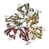
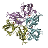
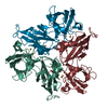
| Title | Crystal Structure of Two-Domain Laccase mutant I170F from Streptomyces griseoflavus | |||||||||
 Components Components | Two-domain laccase | |||||||||
 Keywords Keywords |  OXIDOREDUCTASE / Two-Domain Laccase / laccase / Streptomyces griseoflavus OXIDOREDUCTASE / Two-Domain Laccase / laccase / Streptomyces griseoflavus | |||||||||
| Function / homology |  Function and homology information Function and homology informationhydroquinone:oxygen oxidoreductase activity / laccase / copper ion bindingSimilarity search - Function | |||||||||
| Biological species |  Streptomyces griseoflavus (bacteria) Streptomyces griseoflavus (bacteria) | |||||||||
| Method | X-RAY DIFFRACTION / SYNCHROTRON / MOLECULAR REPLACEMENT / Resolution: 1.85 Å | |||||||||
 Authors Authors | Gabdulkhakov, A.G. / Tishchenko, T.V. / Kolyadenko, I.A. | |||||||||
| Funding support |  Russian Federation, 2items Russian Federation, 2items
| |||||||||
 Citation Citation | Journal: Int J Mol Sci / Year: 2019 Title: Investigations of Accessibility of T2/T3 Copper Center of Two-Domain Laccase fromStreptomyces griseoflavusAc-993. Authors: Gabdulkhakov, A. / Kolyadenko, I. / Kostareva, O. / Mikhaylina, A. / Oliveira, P. / Tamagnini, P. / Lisov, A. / Tishchenko, S. | |||||||||
| History |
|
- Structure visualization
| Structure viewer | Molecule: MolmilJmol/JSmol |
|---|
- Downloads & links
-Download
| PDBx/mmCIF format | 6rh9.cif.gz | 1.3 MB | Display | PDBx/mmCIF format |
|---|---|---|---|---|
| PDB format | pdb6rh9.ent.gz | 1.1 MB | Display | PDB format |
| PDBx/mmJSON format | 6rh9.json.gz | Tree view | PDBx/mmJSON format | |
| Others |  Other downloads Other downloads |
-Validation report
| Arichive directory | https://data.pdbj.org/pub/pdb/validation_reports/rh/6rh9ftp://data.pdbj.org/pub/pdb/validation_reports/rh/6rh9 | HTTPS FTP |
|---|
-Related structure data
| Related structure data | 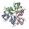 5mkmC  6fc7C  6fdjC  6rhqC  6s0oC  5o4qS C: citing same article ( S: Starting model for refinement |
|---|---|
| Similar structure data |


-Links
 PDBj
PDBj


- Assembly
| Deposited unit | 
| ||||||||
|---|---|---|---|---|---|---|---|---|---|
| 1 | 
| ||||||||
| 2 | 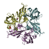
| ||||||||
| 3 | 
| ||||||||
| 4 | 
| ||||||||
| Unit cell |
|
-Components
-Protein , 1 types, 12 molecules ABCDEFGHIJKL
| #1: Protein | Mass: 34819.668 Da / Num. of mol.: 12 / Mutation: I170F Source method: isolated from a genetically manipulated source Source: (gene. exp.) Streptomyces griseoflavus (bacteria) / Production host: Escherichia coli (E. coli) / References: UniProt: A0A0M4FJ81, laccase |
|---|
-Non-polymers , 6 types, 529 molecules 


| #2: Chemical | ChemComp-CU / Copper Mass: 63.546 Da / Num. of mol.: 48 / Source method: obtained synthetically / Formula: Cu Mass: 63.546 Da / Num. of mol.: 48 / Source method: obtained synthetically / Formula: Cu#3: Chemical | ChemComp-EDO / Ethylene glycol Mass: 62.068 Da / Num. of mol.: 7 / Source method: obtained synthetically / Formula: C2H6O2 Mass: 62.068 Da / Num. of mol.: 7 / Source method: obtained synthetically / Formula: C2H6O2#4: Chemical | ChemComp-SO4 / | Sulfate Mass: 96.063 Da / Num. of mol.: 1 / Source method: obtained synthetically / Formula: SO4 Mass: 96.063 Da / Num. of mol.: 1 / Source method: obtained synthetically / Formula: SO4#5: Chemical | ChemComp-GOL / | Glycerol Mass: 92.094 Da / Num. of mol.: 1 / Source method: obtained synthetically / Formula: C3H8O3 Mass: 92.094 Da / Num. of mol.: 1 / Source method: obtained synthetically / Formula: C3H8O3#6: Chemical | ChemComp-OXY / | Oxygen Mass: 31.999 Da / Num. of mol.: 1 / Source method: obtained synthetically / Formula: O2 Mass: 31.999 Da / Num. of mol.: 1 / Source method: obtained synthetically / Formula: O2#7: Water | ChemComp-HOH / | WaterMass: 18.015 Da / Num. of mol.: 471 / Source method: isolated from a natural source / Formula: H2O |
|---|
-Experimental details
-Experiment
| Experiment | Method: X-RAY DIFFRACTION / Number of used crystals: 1 |
|---|
- Sample preparation
| Crystal | Density Matthews: 2.34 Å3/Da / Density % sol: 47.44 % |
|---|---|
| Crystal grow | Temperature: 296 K / Method: vapor diffusion, hanging drop / pH: 9 Details: 25% Smear medium PEG, 0.1 M Bicine, 0.1 M NaCl, 0.05M H3BO3-NaOH |
-Data collection
| Diffraction | Mean temperature: 100 K / Serial crystal experiment: N |
|---|---|
| Diffraction source | Source: SYNCHROTRON / Site: BESSY  / Beamline: 14.1 / Wavelength: 0.97242 Å / Beamline: 14.1 / Wavelength: 0.97242 Å |
| Detector | Type: DECTRIS PILATUS3 S 6M / Detector: PIXEL / Date: Oct 27, 2017 |
| Radiation | Protocol: SINGLE WAVELENGTH / Monochromatic (M) / Laue (L): M / Scattering type: x-ray |
| Radiation wavelength | Wavelength: 0.97242 Å / Relative weight: 1 |
| Reflection | Resolution: 1.85→50 Å / Num. obs: 256815 / % possible obs: 91.4 % / Redundancy: 2.08 % / Rrim(I) all: 0.109 / Net I/σ(I): 10.9 |
| Reflection shell | Resolution: 1.85→1.9 Å / Rmerge(I) obs: 0.8 / Num. unique obs: 12984 |
- Processing
| Software |
| |||||||||||||||||||||||||||||||||||||||||||||||||||||||||||||||||||||||||||||||||||||||||||||||||||||||||||||||||||||||||||||||||||||||||||||||||||||||||||||||||||||||||||||||||||||||||||||||||||||||||||||||||||||||||
|---|---|---|---|---|---|---|---|---|---|---|---|---|---|---|---|---|---|---|---|---|---|---|---|---|---|---|---|---|---|---|---|---|---|---|---|---|---|---|---|---|---|---|---|---|---|---|---|---|---|---|---|---|---|---|---|---|---|---|---|---|---|---|---|---|---|---|---|---|---|---|---|---|---|---|---|---|---|---|---|---|---|---|---|---|---|---|---|---|---|---|---|---|---|---|---|---|---|---|---|---|---|---|---|---|---|---|---|---|---|---|---|---|---|---|---|---|---|---|---|---|---|---|---|---|---|---|---|---|---|---|---|---|---|---|---|---|---|---|---|---|---|---|---|---|---|---|---|---|---|---|---|---|---|---|---|---|---|---|---|---|---|---|---|---|---|---|---|---|---|---|---|---|---|---|---|---|---|---|---|---|---|---|---|---|---|---|---|---|---|---|---|---|---|---|---|---|---|---|---|---|---|---|---|---|---|---|---|---|---|---|---|---|---|---|---|---|---|---|
| Refinement | Method to determine structure: MOLECULAR REPLACEMENT Starting model: 5O4Q Resolution: 1.85→47.337 Å / SU ML: 0.32 / Cross valid method: FREE R-VALUE / σ(F): 1.95 / Phase error: 28.66
| |||||||||||||||||||||||||||||||||||||||||||||||||||||||||||||||||||||||||||||||||||||||||||||||||||||||||||||||||||||||||||||||||||||||||||||||||||||||||||||||||||||||||||||||||||||||||||||||||||||||||||||||||||||||||
| Solvent computation | Shrinkage radii: 0.9 Å / VDW probe radii: 1.11 Å | |||||||||||||||||||||||||||||||||||||||||||||||||||||||||||||||||||||||||||||||||||||||||||||||||||||||||||||||||||||||||||||||||||||||||||||||||||||||||||||||||||||||||||||||||||||||||||||||||||||||||||||||||||||||||
| Refinement step | Cycle: LAST / Resolution: 1.85→47.337 Å
| |||||||||||||||||||||||||||||||||||||||||||||||||||||||||||||||||||||||||||||||||||||||||||||||||||||||||||||||||||||||||||||||||||||||||||||||||||||||||||||||||||||||||||||||||||||||||||||||||||||||||||||||||||||||||
| Refine LS restraints |
| |||||||||||||||||||||||||||||||||||||||||||||||||||||||||||||||||||||||||||||||||||||||||||||||||||||||||||||||||||||||||||||||||||||||||||||||||||||||||||||||||||||||||||||||||||||||||||||||||||||||||||||||||||||||||
| LS refinement shell |
|
