 Movie
Movie Controller

[English] 日本語
 Yorodumi
- PDB-3kvd: Crystal structure of the Neisseria meningitidis Factor H binding ... -
+ Open data

- Basic information
| Entry | Database: PDB / ID: 3kvd | ||||||
|---|---|---|---|---|---|---|---|
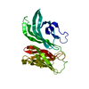
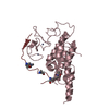
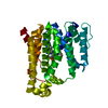
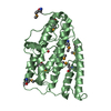
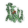
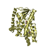
| Title | Crystal structure of the Neisseria meningitidis Factor H binding protein, fHbp (GNA1870) at 2.0 A resolution | ||||||
 Components Components | Lipoprotein | ||||||
 Keywords Keywords | PROTEIN BINDING / alternative complement pathway / antigen / meningococcal vaccines / Lipoprotein | ||||||
| Function / homology |  Function and homology information Function and homology information | ||||||
| Biological species |  Neisseria meningitidis (bacteria) Neisseria meningitidis (bacteria) | ||||||
| Method | X-RAY DIFFRACTION / SYNCHROTRON / MOLECULAR REPLACEMENT / Resolution: 2 Å | ||||||
 Authors Authors | Cendron, L. / Veggi, D. / Girardi, E. / Zanotti, G. | ||||||
 Citation Citation | Journal: Acta Crystallogr.,Sect.F / Year: 2011 Title: Structure of the uncomplexed Neisseria meningitidis factor H-binding protein fHbp (rLP2086). Authors: Cendron, L. / Veggi, D. / Girardi, E. / Zanotti, G. #1: Journal: Nature / Year: 2009Title: Neisseria Meningitidis Recruits Factor H using Protein Mimicry of Host Carbohydrates Authors: Schneider, M.C. / Prosser, B.E. / Caesar, J.J. / Kugelberg, E. / Li, S. / Zhang, Q. / Quoraishi, S. / Lovett, J.E. / Deane, J.E. / Sim, R.B. / Roversi, P. / Johnson, S. / Tang, C.M. / Lea, S.M. | ||||||
| History |
|
- Structure visualization
| Structure viewer | Molecule: MolmilJmol/JSmol |
|---|
- Downloads & links
-Download
| PDBx/mmCIF format | 3kvd.cif.gz | 62.6 KB | Display | PDBx/mmCIF format |
|---|---|---|---|---|
| PDB format | pdb3kvd.ent.gz | 45.6 KB | Display | PDB format |
| PDBx/mmJSON format | 3kvd.json.gz | Tree view | PDBx/mmJSON format | |
| Others |  Other downloads Other downloads |
-Validation report
| Arichive directory | https://data.pdbj.org/pub/pdb/validation_reports/kv/3kvdftp://data.pdbj.org/pub/pdb/validation_reports/kv/3kvd | HTTPS FTP |
|---|
-Related structure data
| Related structure data |  2w80S S: Starting model for refinement |
|---|---|
| Similar structure data |


-Links
 PDBj
PDBj


- Assembly
| Deposited unit | 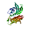
| ||||||||
|---|---|---|---|---|---|---|---|---|---|
| 1 |
| ||||||||
| Unit cell |
|
-Components
| #1: Protein | / Lipoprotein GNA1870 Mass: 25971.002 Da / Num. of mol.: 1 / Fragment: UNP RESIDUES 33-274 Source method: isolated from a genetically manipulated source Source: (gene. exp.) Neisseria meningitidis (bacteria) / Strain: C58 / Gene: gna1870 / Production host: Escherichia coli (E. coli) / References: UniProt: Q6QCC2 |
|---|---|
| #2: Water | ChemComp-HOH / Water Mass: 18.015 Da / Num. of mol.: 189 / Source method: isolated from a natural source / Formula: H2O Mass: 18.015 Da / Num. of mol.: 189 / Source method: isolated from a natural source / Formula: H2O |
-Experimental details
-Experiment
| Experiment | Method: X-RAY DIFFRACTION / Number of used crystals: 1 |
|---|
- Sample preparation
| Crystal | Density Matthews: 2.74 Å3/Da / Density % sol: 55.05 % |
|---|---|
| Crystal grow | Temperature: 293 K / Method: vapor diffusion / pH: 7.5 Details: 25% PEG 2000 MME, 300 mM Na acetate, 100 mM Tris, pH 7.5, VAPOR DIFFUSION, temperature 293K |
-Data collection
| Diffraction | Mean temperature: 100 K |
|---|---|
| Diffraction source | Source: SYNCHROTRON / Site: ELETTRA  / Beamline: 5.2R / Wavelength: 1.2 Å / Beamline: 5.2R / Wavelength: 1.2 Å |
| Detector | Type: MAR scanner 345 mm plate / Detector: IMAGE PLATE / Date: Feb 22, 2004 |
| Radiation | Monochromator: Si(111) / Protocol: SINGLE WAVELENGTH / Monochromatic (M) / Laue (L): M / Scattering type: x-ray |
| Radiation wavelength | Wavelength: 1.2 Å / Relative weight: 1 |
| Reflection | Resolution: 2→60 Å / Num. all: 17912 / Num. obs: 17912 / % possible obs: 98 % / Observed criterion σ(F): 0 / Observed criterion σ(I): 0 / Redundancy: 3.9 % / Biso Wilson estimate: 30.6 Å2 / Rmerge(I) obs: 0.22 / Net I/σ(I): 6.6 |
| Reflection shell | Resolution: 2→2.2 Å / Redundancy: 3.6 % / Rmerge(I) obs: 0.47 / Mean I/σ(I) obs: 1.8 / Num. unique all: 2010 / % possible all: 90.7 |
- Processing
| Software |
| ||||||||||||||||||||||||||||||||||||||||||||||||||||||||||||||||||||||||||||||||||||||||||||||||||||||||||||||||||||||||||||||||||||||||||||||||||||||||||||||||||||||||||
|---|---|---|---|---|---|---|---|---|---|---|---|---|---|---|---|---|---|---|---|---|---|---|---|---|---|---|---|---|---|---|---|---|---|---|---|---|---|---|---|---|---|---|---|---|---|---|---|---|---|---|---|---|---|---|---|---|---|---|---|---|---|---|---|---|---|---|---|---|---|---|---|---|---|---|---|---|---|---|---|---|---|---|---|---|---|---|---|---|---|---|---|---|---|---|---|---|---|---|---|---|---|---|---|---|---|---|---|---|---|---|---|---|---|---|---|---|---|---|---|---|---|---|---|---|---|---|---|---|---|---|---|---|---|---|---|---|---|---|---|---|---|---|---|---|---|---|---|---|---|---|---|---|---|---|---|---|---|---|---|---|---|---|---|---|---|---|---|---|---|---|---|
| Refinement | Method to determine structure: MOLECULAR REPLACEMENT Starting model: 2W80 Resolution: 2→60 Å / Cor.coef. Fo:Fc: 0.932 / Cor.coef. Fo:Fc free: 0.925 / SU B: 4.945 / SU ML: 0.134 / Cross valid method: THROUGHOUT / ESU R: 0.222 / ESU R Free: 0.178 / Stereochemistry target values: MAXIMUM LIKELIHOOD / Details: HYDROGENS HAVE BEEN ADDED IN THE RIDING POSITIONS
| ||||||||||||||||||||||||||||||||||||||||||||||||||||||||||||||||||||||||||||||||||||||||||||||||||||||||||||||||||||||||||||||||||||||||||||||||||||||||||||||||||||||||||
| Solvent computation | Ion probe radii: 0.8 Å / Shrinkage radii: 0.8 Å / VDW probe radii: 1.4 Å / Solvent model: MASK | ||||||||||||||||||||||||||||||||||||||||||||||||||||||||||||||||||||||||||||||||||||||||||||||||||||||||||||||||||||||||||||||||||||||||||||||||||||||||||||||||||||||||||
| Displacement parameters | Biso mean: 30.583 Å2
| ||||||||||||||||||||||||||||||||||||||||||||||||||||||||||||||||||||||||||||||||||||||||||||||||||||||||||||||||||||||||||||||||||||||||||||||||||||||||||||||||||||||||||
| Refinement step | Cycle: LAST / Resolution: 2→60 Å
| ||||||||||||||||||||||||||||||||||||||||||||||||||||||||||||||||||||||||||||||||||||||||||||||||||||||||||||||||||||||||||||||||||||||||||||||||||||||||||||||||||||||||||
| Refine LS restraints |
| ||||||||||||||||||||||||||||||||||||||||||||||||||||||||||||||||||||||||||||||||||||||||||||||||||||||||||||||||||||||||||||||||||||||||||||||||||||||||||||||||||||||||||
| LS refinement shell | Resolution: 2→2.052 Å / Total num. of bins used: 20
|
