 Movie
Movie Controller

[English] 日本語
 Yorodumi
- PDB-2y7s: Structure of a designed meningococcal antigen (factor H binding p... -
+ Open data

- Basic information
| Entry | Database: PDB / ID: 2y7s | ||||||
|---|---|---|---|---|---|---|---|
| Title | Structure of a designed meningococcal antigen (factor H binding protein, mutant G1) inducing broad protective immunity | ||||||
 Components Components | FACTOR H BINDING PROTEIN | ||||||
 Keywords Keywords |  IMMUNE SYSTEM / ANTIGEN / EPITOPE / ANTIBODY / STRUCTURE-BASED DESIGN / VACCINE IMMUNE SYSTEM / ANTIGEN / EPITOPE / ANTIBODY / STRUCTURE-BASED DESIGN / VACCINE | ||||||
| Function / homology |  Function and homology information Function and homology information | ||||||
| Biological species |  NEISSERIA MENINGITIDIS SEROGROUP B (bacteria) NEISSERIA MENINGITIDIS SEROGROUP B (bacteria) | ||||||
| Method | X-RAY DIFFRACTION / SYNCHROTRON / MOLECULAR REPLACEMENT / Resolution: 1.9 Å | ||||||
 Authors Authors | Malito, E. / Spraggon, G. / Bottomley, M.J. | ||||||
 Citation Citation | Journal: Sci.Transl.Med / Year: 2011 Title: Rational Design of a Meningococcal Antigen Inducing Broad Protective Immunity. Authors: Scarselli, M. / Arico, B. / Brunelli, B. / Savino, S. / Di Marcello, F. / Palumbo, E. / Veggi, D. / Ciucchi, L. / Cartocci, E. / Bottomley, M.J. / Malito, E. / Lo Surdo, P. / Comanducci, M. ...Authors: Scarselli, M. / Arico, B. / Brunelli, B. / Savino, S. / Di Marcello, F. / Palumbo, E. / Veggi, D. / Ciucchi, L. / Cartocci, E. / Bottomley, M.J. / Malito, E. / Lo Surdo, P. / Comanducci, M. / Giuliani, M.M. / Cantini, F. / Dragonetti, S. / Colaprico, A. / Doro, F. / Giannetti, P. / Pallaoro, M. / Brogioni, B. / Tontini, M. / Hilleringmann, M. / Nardi-Dei, V. / Banci, L. / Pizza, M. / Rappuoli, R. | ||||||
| History |
| ||||||
| Remark 700 | SHEET DETERMINATION METHOD: DSSP THE SHEETS PRESENTED AS "AC" IN EACH CHAIN ON SHEET RECORDS BELOW ... SHEET DETERMINATION METHOD: DSSP THE SHEETS PRESENTED AS "AC" IN EACH CHAIN ON SHEET RECORDS BELOW IS ACTUALLY AN 8-STRANDED BARREL THIS IS REPRESENTED BY A 9-STRANDED SHEET IN WHICH THE FIRST AND LAST STRANDS ARE IDENTICAL. THE SHEETS PRESENTED AS "BC" IN EACH CHAIN ON SHEET RECORDS BELOW IS ACTUALLY AN 8-STRANDED BARREL THIS IS REPRESENTED BY A 9-STRANDED SHEET IN WHICH THE FIRST AND LAST STRANDS ARE IDENTICAL. |
- Structure visualization
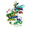
| Structure viewer | Molecule: MolmilJmol/JSmol |
|---|
- Downloads & links
-Download
| PDBx/mmCIF format | 2y7s.cif.gz | 116.2 KB | Display | PDBx/mmCIF format |
|---|---|---|---|---|
| PDB format | pdb2y7s.ent.gz | 89.1 KB | Display | PDB format |
| PDBx/mmJSON format | 2y7s.json.gz | Tree view | PDBx/mmJSON format | |
| Others |  Other downloads Other downloads |
-Validation report
| Arichive directory | https://data.pdbj.org/pub/pdb/validation_reports/y7/2y7sftp://data.pdbj.org/pub/pdb/validation_reports/y7/2y7s | HTTPS FTP |
|---|
-Related structure data
| Related structure data |  2w80S S: Starting model for refinement |
|---|---|
| Similar structure data |


-Links
 PDBj
PDBj


- Assembly
| Deposited unit | 
| ||||||||
|---|---|---|---|---|---|---|---|---|---|
| 1 | 
| ||||||||
| 2 | 
| ||||||||
| Unit cell |
|
-Components
| #1: Protein | Mass: 27718.893 Da / Num. of mol.: 2 / Fragment: RESIDUES 73-320 / Mutation: YES Source method: isolated from a genetically manipulated source Details: THE SEQUENCE OF FHBP CORRESPONDS TO VARIANT 1.1 WITH 23 MUTATIONS FROM VARIANTS 2 AND 3 Source: (gene. exp.) NEISSERIA MENINGITIDIS SEROGROUP B (bacteria)Strain: MC58 / Variant: 1.1 / Production host: ESCHERICHIA COLI (E. coli) / Strain (production host): BL21(DE3) / References: UniProt: Q9JXV4#2: Water | ChemComp-HOH / | Water Mass: 18.015 Da / Num. of mol.: 569 / Source method: isolated from a natural source / Formula: H2O Mass: 18.015 Da / Num. of mol.: 569 / Source method: isolated from a natural source / Formula: H2OCompound details | ENGINEERED RESIDUE IN CHAIN A, ILE 199 TO LEU ENGINEERED RESIDUE IN CHAIN A, ALA 200 TO GLY ...ENGINEERED | |
|---|
-Experimental details
-Experiment
| Experiment | Method: X-RAY DIFFRACTION / Number of used crystals: 1 |
|---|
- Sample preparation
| Crystal | Density Matthews: 2.6 Å3/Da / Density % sol: 53 % / Description: NONE |
|---|---|
| Crystal grow | pH: 6.6 / Details: 20% PEG 3350, 0.2M AMMONIUM FORMATE, PH 6.6 |
-Data collection
| Diffraction | Mean temperature: 100 K |
|---|---|
| Diffraction source | Source: SYNCHROTRON / Site: ALS  / Beamline: 5.0.3 / Wavelength: 0.97 / Beamline: 5.0.3 / Wavelength: 0.97 |
| Detector | Type: ADSC QUANTUM 315r / Detector: CCD / Date: Dec 4, 2010 |
| Radiation | Protocol: SINGLE WAVELENGTH / Monochromatic (M) / Laue (L): M / Scattering type: x-ray |
| Radiation wavelength | Wavelength: 0.97 Å / Relative weight: 1 |
| Reflection | Resolution: 1.9→50 Å / Num. obs: 41959 / % possible obs: 99 % / Observed criterion σ(I): 2 / Redundancy: 3.7 % / Biso Wilson estimate: 11.87 Å2 / Rmerge(I) obs: 0.1 / Net I/σ(I): 9.4 |
| Reflection shell | Resolution: 1.9→2 Å / Redundancy: 3.5 % / Rmerge(I) obs: 0.37 / Mean I/σ(I) obs: 3.5 / % possible all: 99 |
- Processing
| Software |
| ||||||||||||||||||||||||||||||||||||||||||||||||||||||||||||||||||||||||||||||||||||||||||||||||||||||||||||||||
|---|---|---|---|---|---|---|---|---|---|---|---|---|---|---|---|---|---|---|---|---|---|---|---|---|---|---|---|---|---|---|---|---|---|---|---|---|---|---|---|---|---|---|---|---|---|---|---|---|---|---|---|---|---|---|---|---|---|---|---|---|---|---|---|---|---|---|---|---|---|---|---|---|---|---|---|---|---|---|---|---|---|---|---|---|---|---|---|---|---|---|---|---|---|---|---|---|---|---|---|---|---|---|---|---|---|---|---|---|---|---|---|---|---|
| Refinement | Method to determine structure: MOLECULAR REPLACEMENT Starting model: PDB ENTRY 2W80 Resolution: 1.9→49.817 Å / SU ML: 0.24 / σ(F): 1.36 / Phase error: 22.55 / Stereochemistry target values: ML
| ||||||||||||||||||||||||||||||||||||||||||||||||||||||||||||||||||||||||||||||||||||||||||||||||||||||||||||||||
| Solvent computation | Shrinkage radii: 0.9 Å / VDW probe radii: 1.11 Å / Solvent model: FLAT BULK SOLVENT MODEL / Bsol: 22.31 Å2 / ksol: 0.299 e/Å3 | ||||||||||||||||||||||||||||||||||||||||||||||||||||||||||||||||||||||||||||||||||||||||||||||||||||||||||||||||
| Displacement parameters | Biso mean: 14 Å2
| ||||||||||||||||||||||||||||||||||||||||||||||||||||||||||||||||||||||||||||||||||||||||||||||||||||||||||||||||
| Refinement step | Cycle: LAST / Resolution: 1.9→49.817 Å
| ||||||||||||||||||||||||||||||||||||||||||||||||||||||||||||||||||||||||||||||||||||||||||||||||||||||||||||||||
| Refine LS restraints |
| ||||||||||||||||||||||||||||||||||||||||||||||||||||||||||||||||||||||||||||||||||||||||||||||||||||||||||||||||
| LS refinement shell |
|
