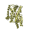
 Movie
Movie Controller


+ Open data

- Basic information
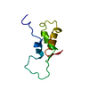
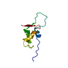
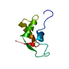
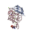
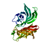
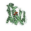
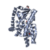
| Entry | Database: PDB / ID: 1hc8 | ||||||
|---|---|---|---|---|---|---|---|
| Title | CRYSTAL STRUCTURE OF A CONSERVED RIBOSOMAL PROTEIN-RNA COMPLEX | ||||||
 Components Components |
| ||||||
 Keywords Keywords |  RIBOSOME / RIBOSOMAL RNA / TERTIARY STRUCTURE / RNA-PROTEIN RIBOSOME / RIBOSOMAL RNA / TERTIARY STRUCTURE / RNA-PROTEIN | ||||||
| Function / homology |  Function and homology information Function and homology informationlarge ribosomal subunit rRNA binding / ribosome / structural constituent of ribosome / ribonucleoprotein complex / translation / DNA bindingSimilarity search - Function | ||||||
| Biological species |   BACILLUS STEAROTHERMOPHILUS (bacteria)ESCHERICHIA COLI (E. coli) BACILLUS STEAROTHERMOPHILUS (bacteria)ESCHERICHIA COLI (E. coli) | ||||||
| Method | X-RAY DIFFRACTION / SYNCHROTRON / MAD / Resolution: 2.8 Å | ||||||
 Authors Authors | Conn, G.L. / Draper, D.E. / Lattman, E.E. / Gittis, A.G. | ||||||
 Citation Citation | Journal: J.Mol.Biol. / Year: 2002 Title: A Compact RNA Tertiary Structure Contains a Buried Backbone-K+ Complex Authors: Conn, G.L. / Gittis, A.G. / Lattman, E.E. / Misra, V.K. / Draper, D.E. #1: Journal: Science / Year: 1999Title: Crystal Structure of a Conserved Ribosomal Protein-RNA Complex Authors: Conn, G.L. / Draper, D.E. / Lattman, E.E. / Gittis, A.G. | ||||||
| History |
|
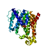
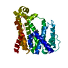
- Structure visualization
| Structure viewer | Molecule: MolmilJmol/JSmol |
|---|
- Downloads & links
-Download
| PDBx/mmCIF format | 1hc8.cif.gz | 105.5 KB | Display | PDBx/mmCIF format |
|---|---|---|---|---|
| PDB format | pdb1hc8.ent.gz | 78.3 KB | Display | PDB format |
| PDBx/mmJSON format | 1hc8.json.gz | Tree view | PDBx/mmJSON format | |
| Others |  Other downloads Other downloads |
-Validation report
| Arichive directory | https://data.pdbj.org/pub/pdb/validation_reports/hc/1hc8ftp://data.pdbj.org/pub/pdb/validation_reports/hc/1hc8 | HTTPS FTP |
|---|
-Related structure data
| Related structure data | |
|---|---|
| Similar structure data |


-Links
 PDBj
PDBj


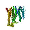
- Assembly
| Deposited unit | 
| ||||||||
|---|---|---|---|---|---|---|---|---|---|
| 1 | 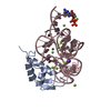
| ||||||||
| 2 | 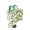
| ||||||||
| Unit cell |
| ||||||||
| Noncrystallographic symmetry (NCS) | NCS oper: (Code: given Matrix: (0.043157, -0.998994, -0.012207), Vector : Details | COMPLEX OF THE RIBOSOMAL PROTEIN L11 AND THERIBOSOMAL 23S RNA FRAGMENT | |
-Components
-Protein / RNA chain , 2 types, 4 molecules ABCD
| #1: Protein | Mass: 8167.547 Da / Num. of mol.: 2 / Fragment: C-TERMINAL DOMAIN OF RIBOSOMAL PROTEIN L11 Source method: isolated from a genetically manipulated source Source: (gene. exp.) BACILLUS STEAROTHERMOPHILUS (bacteria) / Plasmid: PET11 / Production host: ESCHERICHIA COLI (E. coli) / Strain (production host): BL21(DE3) / References: UniProt: P56210#2: RNA chain | Mass: 18885.150 Da / Num. of mol.: 2 / Fragment: NTS 1051-1108 FROM E. COLI 23S RRNA / Mutation: YES Source method: isolated from a genetically manipulated source Source: (gene. exp.) ESCHERICHIA COLI (E. coli)Description: RNA SYNTHESIZED BY IN VITRO TRANSCRIPTION USING T7 RNA POLYMERASE Production host: ESCHERICHIA COLI (E. coli) |
|---|
-Non-polymers , 4 types, 40 molecules 


| #3: Chemical | ChemComp-MG /  Mass: 24.305 Da / Num. of mol.: 21 / Source method: obtained synthetically / Formula: Mg Mass: 24.305 Da / Num. of mol.: 21 / Source method: obtained synthetically / Formula: Mg#4: Chemical |  Mass: 39.098 Da / Num. of mol.: 2 / Source method: obtained synthetically / Formula: K Mass: 39.098 Da / Num. of mol.: 2 / Source method: obtained synthetically / Formula: K#5: Chemical | ChemComp-OS / Osmium Mass: 190.230 Da / Num. of mol.: 4 / Source method: obtained synthetically / Formula: Os Mass: 190.230 Da / Num. of mol.: 4 / Source method: obtained synthetically / Formula: Os#6: Water | ChemComp-HOH / | WaterMass: 18.015 Da / Num. of mol.: 13 / Source method: isolated from a natural source / Formula: H2O |
|---|
-Details
| Compound details | CHAIN C, D ENGINEERED |
|---|
-Experimental details
-Experiment
| Experiment | Method: X-RAY DIFFRACTION / Number of used crystals: 1 |
|---|
- Sample preparation
| Crystal | Density Matthews: 3.35 Å3/Da / Density % sol: 70 % | ||||||||||||||||||||||||||||||||||||
|---|---|---|---|---|---|---|---|---|---|---|---|---|---|---|---|---|---|---|---|---|---|---|---|---|---|---|---|---|---|---|---|---|---|---|---|---|---|
| Crystal grow | pH: 6.5 Details: PEG 600, MAGNESIUM ACETATE, COBALT HEXAMINE CHLORIDE, SODIUM CACODYLATE, KCL, pH 6.50 | ||||||||||||||||||||||||||||||||||||
| Crystal grow | *PLUS Temperature: 37 ℃ / pH: 6.5 / Method: vapor diffusion, sitting drop / Details: Conn, G.L., (1999) Science, 284, 1171. | ||||||||||||||||||||||||||||||||||||
| Components of the solutions | *PLUS
|
-Data collection
| Diffraction | Mean temperature: 100 K |
|---|---|
| Diffraction source | Source: SYNCHROTRON / Site: NSLS  / Beamline: X4A / Wavelength: 1.13951 / Beamline: X4A / Wavelength: 1.13951 |
| Detector | Type: RIGAKU IMAGE PLATE / Detector: IMAGE PLATE / Date: Sep 9, 1998 |
| Radiation | Protocol: SINGLE WAVELENGTH / Monochromatic (M) / Laue (L): M / Scattering type: x-ray |
| Radiation wavelength | Wavelength: 1.13951 Å / Relative weight: 1 |
| Reflection | Resolution: 2.8→20 Å / Num. obs: 17542 / % possible obs: 94.5 % / Observed criterion σ(I): 0 / Redundancy: 5 % / Biso Wilson estimate: 75.5 Å2 / Rmerge(I) obs: 0.24 / Net I/σ(I): 5.9 |
| Reflection shell | Resolution: 2.8→2.93 Å / Redundancy: 4 % / Rmerge(I) obs: 0.27 / % possible all: 88.7 |
| Reflection | *PLUS Highest resolution: 2.8 Å / Lowest resolution: 20 Å / Num. measured all: 89078 / Rmerge(I) obs: 0.55 |
| Reflection shell | *PLUS % possible obs: 88.7 % / Rmerge(I) obs: 0.24 / Mean I/σ(I) obs: 5.9 |
- Processing
| Software |
| ||||||||||||||||||||||||||||||||||||||||||||||||||||||||||||
|---|---|---|---|---|---|---|---|---|---|---|---|---|---|---|---|---|---|---|---|---|---|---|---|---|---|---|---|---|---|---|---|---|---|---|---|---|---|---|---|---|---|---|---|---|---|---|---|---|---|---|---|---|---|---|---|---|---|---|---|---|---|
| Refinement | Method to determine structure: MAD / Resolution: 2.8→20 Å / Data cutoff high absF: 10000 / Cross valid method: THROUGHOUT / σ(F): 0 Stereochemistry target values: MAXIMUM LIKELIHOOD USING AMPLITUDES
| ||||||||||||||||||||||||||||||||||||||||||||||||||||||||||||
| Solvent computation | Solvent model: CNS | ||||||||||||||||||||||||||||||||||||||||||||||||||||||||||||
| Refinement step | Cycle: LAST / Resolution: 2.8→20 Å
| ||||||||||||||||||||||||||||||||||||||||||||||||||||||||||||
| Refine LS restraints |
| ||||||||||||||||||||||||||||||||||||||||||||||||||||||||||||
| LS refinement shell | Resolution: 2.8→2.84 Å / Total num. of bins used: 27
| ||||||||||||||||||||||||||||||||||||||||||||||||||||||||||||
| Xplor file |
| ||||||||||||||||||||||||||||||||||||||||||||||||||||||||||||
| LS refinement shell | *PLUS Rfactor obs: 0.368 |
