 Movie
Movie Controller

[English] 日本語
 Yorodumi
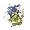
- PDB-3ko4: Crystal structure of D-Tyr-tRNA(Tyr) deacylase from Plasmodium fa... -
+ Open data

- Basic information
| Entry | Database: PDB / ID: 3ko4 | ||||||
|---|---|---|---|---|---|---|---|
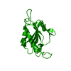
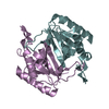
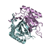
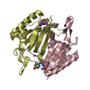
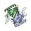
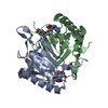
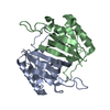
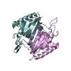
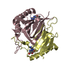
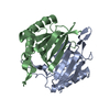
| Title | Crystal structure of D-Tyr-tRNA(Tyr) deacylase from Plasmodium falciparum in complex with ADP | ||||||
 Components Components | D-tyrosyl-tRNA(Tyr) deacylase | ||||||
 Keywords Keywords |  HYDROLASE / DTD / Deacylase / ADP HYDROLASE / DTD / Deacylase / ADP | ||||||
| Function / homology |  Function and homology information Function and homology informationGly-tRNA(Ala) hydrolase activity / D-tyrosyl-tRNA(Tyr) deacylase activity / D-aminoacyl-tRNA deacylase / tRNA metabolic process / tRNA binding / nucleotide binding / cytoplasmSimilarity search - Function | ||||||
| Biological species |  Plasmodium falciparum (malaria parasite P. falciparum) Plasmodium falciparum (malaria parasite P. falciparum) | ||||||
| Method | X-RAY DIFFRACTION / SYNCHROTRON / MOLECULAR REPLACEMENT / molecular replacement / Resolution: 2.7 Å | ||||||
 Authors Authors | Manickam, Y. / Bhatt, T.K. / Sharma, A. | ||||||
 Citation Citation | Journal: J.Biol.Chem. / Year: 2010 Title: Ligand-bound Structures Provide Atomic Snapshots for the Catalytic Mechanism of D-Amino Acid Deacylase Authors: Bhatt, T.K. / Yogavel, M. / Wydau, S. / Berwal, R. / Sharma, A. | ||||||
| History |
|
- Structure visualization
| Structure viewer | Molecule: MolmilJmol/JSmol |
|---|
- Downloads & links
-Download
| PDBx/mmCIF format | 3ko4.cif.gz | 190.2 KB | Display | PDBx/mmCIF format |
|---|---|---|---|---|
| PDB format | pdb3ko4.ent.gz | 153.4 KB | Display | PDB format |
| PDBx/mmJSON format | 3ko4.json.gz | Tree view | PDBx/mmJSON format | |
| Others |  Other downloads Other downloads |
-Validation report
| Arichive directory | https://data.pdbj.org/pub/pdb/validation_reports/ko/3ko4ftp://data.pdbj.org/pub/pdb/validation_reports/ko/3ko4 | HTTPS FTP |
|---|
-Related structure data
| Related structure data |  3knfC 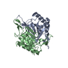 3knpC  3ko3C 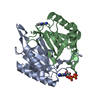 3ko5C 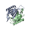 3ko7C 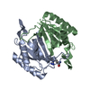 3ko9C 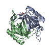 3kobC 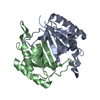 3kocC 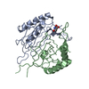 3kodC C: citing same article ( |
|---|---|
| Similar structure data |

-Links
 PDBj
PDBj- Assembly
| Deposited unit | 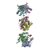
| ||||||||
|---|---|---|---|---|---|---|---|---|---|
| 1 | 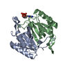
| ||||||||
| 2 | 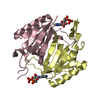
| ||||||||
| 3 | 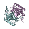
| ||||||||
| Unit cell |
|
-Components
| #1: Protein | Mass: 19233.084 Da / Num. of mol.: 6 Source method: isolated from a genetically manipulated source Source: (gene. exp.) Plasmodium falciparum (malaria parasite P. falciparum)Strain: 3D7 / Gene: dtd / Plasmid: pET28a / Production host:  Escherichia coli (E. coli) / Strain (production host): B834(DE3) Escherichia coli (E. coli) / Strain (production host): B834(DE3)References: UniProt: Q8IIS0, Hydrolases; Acting on ester bonds#2: Chemical | Adenosine diphosphate  Mass: 427.201 Da / Num. of mol.: 3 / Source method: obtained synthetically / Formula: C10H15N5O10P2 / Comment: ADP, energy-carrying molecule*YM Mass: 427.201 Da / Num. of mol.: 3 / Source method: obtained synthetically / Formula: C10H15N5O10P2 / Comment: ADP, energy-carrying molecule*YM#3: Water | ChemComp-HOH / | Water Mass: 18.015 Da / Num. of mol.: 10 / Source method: isolated from a natural source / Formula: H2O Mass: 18.015 Da / Num. of mol.: 10 / Source method: isolated from a natural source / Formula: H2O |
|---|
-Experimental details
-Experiment
| Experiment | Method: X-RAY DIFFRACTION / Number of used crystals: 1 |
|---|
- Sample preparation
| Crystal | Density Matthews: 1.97 Å3/Da / Density % sol: 37.65 % / Mosaicity: 0.943 ° |
|---|---|
| Crystal grow | Temperature: 293 K / Method: vapor diffusion, hanging drop Details: 25% PEG 3350. 0.1 MES , pH 6.2-6.5, VAPOR DIFFUSION, HANGING DROP, temperature 293K PH range: 6.2-6.5 |
-Data collection
| Diffraction | Mean temperature: 100 K |
|---|---|
| Diffraction source | Source: SYNCHROTRON / Site: ESRF  / Beamline: BM14 / Wavelength: 0.97 Å / Beamline: BM14 / Wavelength: 0.97 Å |
| Detector | Type: MARMOSAIC 225 mm CCD / Detector: CCD / Date: Dec 13, 2007 |
| Radiation | Protocol: SINGLE WAVELENGTH / Monochromatic (M) / Laue (L): M / Scattering type: x-ray |
| Radiation wavelength | Wavelength: 0.97 Å / Relative weight: 1 |
| Reflection | Resolution: 2.7→25 Å / Num. obs: 19638 / % possible obs: 81.3 % / Redundancy: 2.7 % / Rmerge(I) obs: 0.08 / Χ2: 0.876 / Net I/σ(I): 6.9 |
| Reflection shell | Resolution: 2.7→2.8 Å / Redundancy: 2.4 % / Rmerge(I) obs: 0.387 / Num. unique all: 1080 / Χ2: 0.729 / % possible all: 44.9 |
-Phasing
| Phasing | Method: molecular replacement |
|---|
- Processing
| Software |
| ||||||||||||||||||||||||||||||||||||||||||||||||||||||||||||||||||||||||||||||||||||||||||
|---|---|---|---|---|---|---|---|---|---|---|---|---|---|---|---|---|---|---|---|---|---|---|---|---|---|---|---|---|---|---|---|---|---|---|---|---|---|---|---|---|---|---|---|---|---|---|---|---|---|---|---|---|---|---|---|---|---|---|---|---|---|---|---|---|---|---|---|---|---|---|---|---|---|---|---|---|---|---|---|---|---|---|---|---|---|---|---|---|---|---|---|
| Refinement | Method to determine structure: MOLECULAR REPLACEMENT Starting model: PfDTD-iodide model (not deposited) Resolution: 2.7→20 Å / Cor.coef. Fo:Fc: 0.932 / Cor.coef. Fo:Fc free: 0.874 / Occupancy max: 1 / Occupancy min: 0.4 / SU B: 19.624 / SU ML: 0.408 / Cross valid method: THROUGHOUT / σ(F): 0 / ESU R Free: 0.536 / Stereochemistry target values: MAXIMUM LIKELIHOOD / Details: HYDROGENS HAVE BEEN ADDED IN THE RIDING POSITIONS
| ||||||||||||||||||||||||||||||||||||||||||||||||||||||||||||||||||||||||||||||||||||||||||
| Solvent computation | Ion probe radii: 0.8 Å / Shrinkage radii: 0.8 Å / VDW probe radii: 1.2 Å / Solvent model: MASK | ||||||||||||||||||||||||||||||||||||||||||||||||||||||||||||||||||||||||||||||||||||||||||
| Displacement parameters | Biso max: 96.17 Å2 / Biso mean: 57.401 Å2 / Biso min: 29.97 Å2
| ||||||||||||||||||||||||||||||||||||||||||||||||||||||||||||||||||||||||||||||||||||||||||
| Refinement step | Cycle: LAST / Resolution: 2.7→20 Å
| ||||||||||||||||||||||||||||||||||||||||||||||||||||||||||||||||||||||||||||||||||||||||||
| Refine LS restraints |
| ||||||||||||||||||||||||||||||||||||||||||||||||||||||||||||||||||||||||||||||||||||||||||
| LS refinement shell | Resolution: 2.7→2.769 Å / Total num. of bins used: 20
|
