 Movie
Movie Controller

[English] 日本語
 Yorodumi
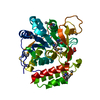
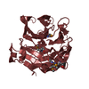
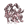
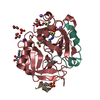
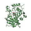
- PDB-2dbo: Crystal structure of D-Tyr-tRNA(Tyr) deacylase from Aquifex aeolicus -
+ Open data

- Basic information
| Entry | Database: PDB / ID: 2dbo | ||||||
|---|---|---|---|---|---|---|---|
| Title | Crystal structure of D-Tyr-tRNA(Tyr) deacylase from Aquifex aeolicus | ||||||
 Components Components | D-tyrosyl-tRNA(Tyr) deacylase | ||||||
 Keywords Keywords |  HYDROLASE / D-amino acid / D-Tyrosine / tRNA / deacylase / Structural Genomics / NPPSFA / National Project on Protein Structural and Functional Analyses / RIKEN Structural Genomics/Proteomics Initiative / RSGI HYDROLASE / D-amino acid / D-Tyrosine / tRNA / deacylase / Structural Genomics / NPPSFA / National Project on Protein Structural and Functional Analyses / RIKEN Structural Genomics/Proteomics Initiative / RSGI | ||||||
| Function / homology |  Function and homology information Function and homology informationSer(Gly)-tRNA(Ala) hydrolase activity / Gly-tRNA(Ala) hydrolase activity / D-tyrosyl-tRNA(Tyr) deacylase activity / D-aminoacyl-tRNA deacylase / tRNA metabolic process / D-amino acid catabolic process / aminoacyl-tRNA editing activity / tRNA binding / cytoplasmSimilarity search - Function | ||||||
| Biological species |   Aquifex aeolicus (bacteria) Aquifex aeolicus (bacteria) | ||||||
| Method | X-RAY DIFFRACTION / SYNCHROTRON / MOLECULAR REPLACEMENT / Resolution: 2.76 Å | ||||||
 Authors Authors | Ishii, T. / Shibata, R. / Bessho, Y. / Shirouzu, M. / Yokoyama, S. / RIKEN Structural Genomics/Proteomics Initiative (RSGI) | ||||||
 Citation Citation | Journal: To be Published Title: Crystal structure of D-Tyr-tRNA(Tyr) deacylase from Aquifex aeolicus Authors: Ishii, T. / Shibata, R. / Bessho, Y. / Shirouzu, M. / Yokoyama, S. | ||||||
| History |
|
- Structure visualization
| Structure viewer | Molecule: MolmilJmol/JSmol |
|---|
- Downloads & links
-Download
| PDBx/mmCIF format | 2dbo.cif.gz | 42.4 KB | Display | PDBx/mmCIF format |
|---|---|---|---|---|
| PDB format | pdb2dbo.ent.gz | 30 KB | Display | PDB format |
| PDBx/mmJSON format | 2dbo.json.gz | Tree view | PDBx/mmJSON format | |
| Others |  Other downloads Other downloads |
-Validation report
| Arichive directory | https://data.pdbj.org/pub/pdb/validation_reports/db/2dboftp://data.pdbj.org/pub/pdb/validation_reports/db/2dbo | HTTPS FTP |
|---|
-Related structure data
| Related structure data | 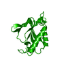 1j7gS S: Starting model for refinement |
|---|---|
| Similar structure data | |
| Other databases |


-Links
 PDBj
PDBj- Assembly
| Deposited unit | 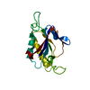
| ||||||||
|---|---|---|---|---|---|---|---|---|---|
| 1 | 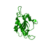
| ||||||||
| Unit cell |
| ||||||||
| Components on special symmetry positions |
|
-Components
| #1: Protein | Mass: 16992.645 Da / Num. of mol.: 1 Source method: isolated from a genetically manipulated source Source: (gene. exp.) Aquifex aeolicus (bacteria) / Strain: VF5 / Plasmid: pET-11b / Production host: Escherichia coli (E. coli)References: UniProt: O66742, Hydrolases; Acting on ester bonds |
|---|---|
| #2: Water | ChemComp-HOH / Water Mass: 18.015 Da / Num. of mol.: 32 / Source method: isolated from a natural source / Formula: H2O Mass: 18.015 Da / Num. of mol.: 32 / Source method: isolated from a natural source / Formula: H2O |
-Experimental details
-Experiment
| Experiment | Method: X-RAY DIFFRACTION / Number of used crystals: 1 |
|---|
- Sample preparation
| Crystal | Density Matthews: 3.28 Å3/Da / Density % sol: 62.52 % |
|---|---|
| Crystal grow | Temperature: 277 K / Method: small tubes / pH: 8 Details: 0.15M sodium chloride, pH 8.0, SMALL TUBES, temperature 277K |
-Data collection
| Diffraction | Mean temperature: 100 K |
|---|---|
| Diffraction source | Source: SYNCHROTRON / Site: Photon Factory  / Beamline: BL-6A / Wavelength: 1 Å / Beamline: BL-6A / Wavelength: 1 Å |
| Detector | Type: ADSC QUANTUM 4 / Detector: CCD / Date: Oct 27, 2005 |
| Radiation | Monochromator: SI(111) / Protocol: SINGLE WAVELENGTH / Monochromatic (M) / Laue (L): M / Scattering type: x-ray |
| Radiation wavelength | Wavelength: 1 Å / Relative weight: 1 |
| Reflection | Resolution: 2.76→38.01 Å / Num. obs: 6373 / % possible obs: 100 % / Redundancy: 12.3 % / Biso Wilson estimate: 46.6 Å2 / Rsym value: 0.09 / Net I/σ(I): 28.8534 |
| Reflection shell | Resolution: 2.76→2.86 Å / Redundancy: 13 % / Mean I/σ(I) obs: 7.72 / Num. unique all: 611 / Rsym value: 0.368 / % possible all: 100 |
- Processing
| Software |
| ||||||||||||||||||||
|---|---|---|---|---|---|---|---|---|---|---|---|---|---|---|---|---|---|---|---|---|---|
| Refinement | Method to determine structure: MOLECULAR REPLACEMENT Starting model: 1J7G Resolution: 2.76→38.01 Å / Rfactor Rfree error: 0.011 / Data cutoff high absF: 111177.44 / Data cutoff low absF: 0 / Isotropic thermal model: RESTRAINED / Cross valid method: THROUGHOUT / σ(F): 0
| ||||||||||||||||||||
| Solvent computation | Solvent model: FLAT MODEL / Bsol: 29.9913 Å2 / ksol: 0.366911 e/Å3 | ||||||||||||||||||||
| Displacement parameters | Biso mean: 36.7 Å2
| ||||||||||||||||||||
| Refine analyze |
| ||||||||||||||||||||
| Refinement step | Cycle: LAST / Resolution: 2.76→38.01 Å
| ||||||||||||||||||||
| Refine LS restraints |
| ||||||||||||||||||||
| LS refinement shell | Resolution: 2.76→2.93 Å / Rfactor Rfree error: 0.035 / Total num. of bins used: 6
| ||||||||||||||||||||
| Xplor file |
|
