 Movie
Movie Controller

[English] 日本語
 Yorodumi
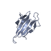
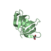
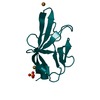
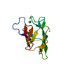
- PDB-3khq: Crystal structure of murine Ig-beta (CD79b) in the monomeric form -
+ Open data

- Basic information
| Entry | Database: PDB / ID: 3khq | ||||||
|---|---|---|---|---|---|---|---|
| Title | Crystal structure of murine Ig-beta (CD79b) in the monomeric form | ||||||
 Components Components | B-cell antigen receptor complex-associated protein beta chain | ||||||
 Keywords Keywords |  PROTEIN BINDING / CD79b / CD79a / Ig-beta / BCR / Ig domain / V-set / Immunoglobulin domain PROTEIN BINDING / CD79b / CD79a / Ig-beta / BCR / Ig domain / V-set / Immunoglobulin domain | ||||||
| Function / homology |  Function and homology information Function and homology informationCD22 mediated BCR regulation / IgM B cell receptor complex / B cell receptor complex / Antigen activates B Cell Receptor (BCR) leading to generation of second messengers / B cell differentiation / B cell receptor signaling pathway / response to bacterium / transmembrane signaling receptor activity / adaptive immune response / external side of plasma membrane ...CD22 mediated BCR regulation / IgM B cell receptor complex / B cell receptor complex / Antigen activates B Cell Receptor (BCR) leading to generation of second messengers / B cell differentiation / B cell receptor signaling pathway / response to bacterium / transmembrane signaling receptor activity / adaptive immune response / external side of plasma membrane / identical protein binding / plasma membraneSimilarity search - Function | ||||||
| Biological species |  Mus musculus (house mouse) Mus musculus (house mouse) | ||||||
| Method | X-RAY DIFFRACTION / SYNCHROTRON / MOLECULAR REPLACEMENT / Resolution: 1.7 Å | ||||||
 Authors Authors | Radaev, S. / Sun, P.D. | ||||||
 Citation Citation | Journal: Structure / Year: 2010 Title: Structural and Functional Studies of Igalphabeta and Its Assembly with the B Cell Antigen Receptor. Authors: Radaev, S. / Zou, Z. / Tolar, P. / Nguyen, K. / Nguyen, A. / Krueger, P.D. / Stutzman, N. / Pierce, S. / Sun, P.D. | ||||||
| History |
|
- Structure visualization
| Structure viewer | Molecule: MolmilJmol/JSmol |
|---|
- Downloads & links
-Download
| PDBx/mmCIF format | 3khq.cif.gz | 32.2 KB | Display | PDBx/mmCIF format |
|---|---|---|---|---|
| PDB format | pdb3khq.ent.gz | 23.8 KB | Display | PDB format |
| PDBx/mmJSON format | 3khq.json.gz | Tree view | PDBx/mmJSON format | |
| Others |  Other downloads Other downloads |
-Validation report
| Arichive directory | https://data.pdbj.org/pub/pdb/validation_reports/kh/3khqftp://data.pdbj.org/pub/pdb/validation_reports/kh/3khq | HTTPS FTP |
|---|
-Related structure data


-Links
 PDBj
PDBj


- Assembly
| Deposited unit | 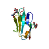
| ||||||||
|---|---|---|---|---|---|---|---|---|---|
| 1 |
| ||||||||
| Unit cell |
|
-Components
| #1: Protein | Mass: 15079.915 Da / Num. of mol.: 1 / Fragment: extracellular domain Source method: isolated from a genetically manipulated source Source: (gene. exp.) Mus musculus (house mouse) / Gene: Cd79b, Igb / Production host:  Escherichia coli (E. coli) / References: UniProt: P15530 Escherichia coli (E. coli) / References: UniProt: P15530 |
|---|---|
| #2: Chemical | ChemComp-MG /   Mass: 24.305 Da / Num. of mol.: 1 / Source method: obtained synthetically / Formula: Mg Mass: 24.305 Da / Num. of mol.: 1 / Source method: obtained synthetically / Formula: Mg |
| #3: Chemical | ChemComp-FLC / Citric acid  Mass: 189.100 Da / Num. of mol.: 1 / Source method: obtained synthetically / Formula: C6H5O7 Mass: 189.100 Da / Num. of mol.: 1 / Source method: obtained synthetically / Formula: C6H5O7 |
| #4: Chemical | ChemComp-GSH / Glutathione  Mass: 307.323 Da / Num. of mol.: 1 / Source method: obtained synthetically / Formula: C10H17N3O6S Mass: 307.323 Da / Num. of mol.: 1 / Source method: obtained synthetically / Formula: C10H17N3O6S |
| #5: Water | ChemComp-HOH / Water Mass: 18.015 Da / Num. of mol.: 58 / Source method: isolated from a natural source / Formula: H2O Mass: 18.015 Da / Num. of mol.: 58 / Source method: isolated from a natural source / Formula: H2O |
-Experimental details
-Experiment
| Experiment | Method: X-RAY DIFFRACTION / Number of used crystals: 1 |
|---|
- Sample preparation
| Crystal | Density Matthews: 1.62 Å3/Da / Density % sol: 23.89 % |
|---|---|
| Crystal grow | Temperature: 298 K / Method: vapor diffusion / pH: 5 Details: 20% Peg 750 MME, 0.2M MgCl2, 100mM Na Citrate, pH 5.0, VAPOR DIFFUSION, temperature 298K |
-Data collection
| Diffraction | Mean temperature: 100 K |
|---|---|
| Diffraction source | Source: SYNCHROTRON / Site: APS  / Beamline: 22-ID / Wavelength: 1 Å / Beamline: 22-ID / Wavelength: 1 Å |
| Detector | Type: MARMOSAIC 300 mm CCD / Detector: CCD / Details: mirrors |
| Radiation | Monochromator: SI(111) / Protocol: SINGLE WAVELENGTH / Monochromatic (M) / Laue (L): M / Scattering type: x-ray |
| Radiation wavelength | Wavelength: 1 Å / Relative weight: 1 |
| Reflection | Resolution: 1.7→50 Å / Num. obs: 10471 / Observed criterion σ(I): -3 / Redundancy: 5.5 % / Biso Wilson estimate: 20.6 Å2 / Rsym value: 0.065 / Net I/σ(I): 21.5 |
| Reflection shell | Resolution: 1.7→1.74 Å / Redundancy: 2.3 % / Mean I/σ(I) obs: 2.6 / Rsym value: 0.317 |
- Processing
| Software |
| ||||||||||||||||||||||||||||||||||||||||||||||||||||||||||||||||||||||||||||||||||||||||||||||||||||||||||||||||||||||||||||||||||||||||||||||||||||||||||||||||||||||||||
|---|---|---|---|---|---|---|---|---|---|---|---|---|---|---|---|---|---|---|---|---|---|---|---|---|---|---|---|---|---|---|---|---|---|---|---|---|---|---|---|---|---|---|---|---|---|---|---|---|---|---|---|---|---|---|---|---|---|---|---|---|---|---|---|---|---|---|---|---|---|---|---|---|---|---|---|---|---|---|---|---|---|---|---|---|---|---|---|---|---|---|---|---|---|---|---|---|---|---|---|---|---|---|---|---|---|---|---|---|---|---|---|---|---|---|---|---|---|---|---|---|---|---|---|---|---|---|---|---|---|---|---|---|---|---|---|---|---|---|---|---|---|---|---|---|---|---|---|---|---|---|---|---|---|---|---|---|---|---|---|---|---|---|---|---|---|---|---|---|---|---|---|
| Refinement | Method to determine structure: MOLECULAR REPLACEMENT / Resolution: 1.7→39.87 Å / Cor.coef. Fo:Fc: 0.964 / Cor.coef. Fo:Fc free: 0.96 / SU B: 2.114 / SU ML: 0.068 / Cross valid method: THROUGHOUT / ESU R: 0.117 / ESU R Free: 0.101 / Stereochemistry target values: MAXIMUM LIKELIHOOD / Details: HYDROGENS HAVE BEEN ADDED IN THE RIDING POSITIONS
| ||||||||||||||||||||||||||||||||||||||||||||||||||||||||||||||||||||||||||||||||||||||||||||||||||||||||||||||||||||||||||||||||||||||||||||||||||||||||||||||||||||||||||
| Solvent computation | Ion probe radii: 0.8 Å / Shrinkage radii: 0.8 Å / VDW probe radii: 1.4 Å / Solvent model: BABINET MODEL WITH MASK | ||||||||||||||||||||||||||||||||||||||||||||||||||||||||||||||||||||||||||||||||||||||||||||||||||||||||||||||||||||||||||||||||||||||||||||||||||||||||||||||||||||||||||
| Displacement parameters | Biso mean: 31.694 Å2
| ||||||||||||||||||||||||||||||||||||||||||||||||||||||||||||||||||||||||||||||||||||||||||||||||||||||||||||||||||||||||||||||||||||||||||||||||||||||||||||||||||||||||||
| Refinement step | Cycle: LAST / Resolution: 1.7→39.87 Å
| ||||||||||||||||||||||||||||||||||||||||||||||||||||||||||||||||||||||||||||||||||||||||||||||||||||||||||||||||||||||||||||||||||||||||||||||||||||||||||||||||||||||||||
| Refine LS restraints |
| ||||||||||||||||||||||||||||||||||||||||||||||||||||||||||||||||||||||||||||||||||||||||||||||||||||||||||||||||||||||||||||||||||||||||||||||||||||||||||||||||||||||||||
| LS refinement shell | Resolution: 1.7→1.744 Å / Total num. of bins used: 20
|
