 Movie
Movie Controller

[English] 日本語
 Yorodumi
- PDB-3kex: Crystal structure of the catalytically inactive kinase domain of ... -
+ Open data

- Basic information
| Entry | Database: PDB / ID: 3kex | ||||||
|---|---|---|---|---|---|---|---|
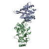
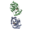
| Title | Crystal structure of the catalytically inactive kinase domain of the human epidermal growth factor receptor 3 (HER3) | ||||||
 Components Components | Receptor tyrosine-protein kinase erbB-3 | ||||||
 Keywords Keywords |  TRANSFERASE / kinase domain / inactive kinase / HER3 / ErbB3 / ATP-binding / Cell membrane / Kinase / Membrane / Nucleotide-binding / Phosphoprotein / Receptor / Tyrosine-protein kinase TRANSFERASE / kinase domain / inactive kinase / HER3 / ErbB3 / ATP-binding / Cell membrane / Kinase / Membrane / Nucleotide-binding / Phosphoprotein / Receptor / Tyrosine-protein kinase | ||||||
| Function / homology |  Function and homology informationneuregulin binding / positive regulation of cardiac muscle tissue development / cranial nerve development / Schwann cell differentiation / neuregulin receptor activity / negative regulation of secretion / endocardial cushion development / ERBB3:ERBB2 complex / GRB7 events in ERBB2 signaling / positive regulation of calcineurin-NFAT signaling cascade ...neuregulin binding / positive regulation of cardiac muscle tissue development / cranial nerve development / Schwann cell differentiation / neuregulin receptor activity / negative regulation of secretion / endocardial cushion development / ERBB3:ERBB2 complex / GRB7 events in ERBB2 signaling / positive regulation of calcineurin-NFAT signaling cascade / peripheral nervous system development / ErbB-3 class receptor binding / negative regulation of cell adhesion / negative regulation of motor neuron apoptotic process / motor neuron apoptotic process / ERBB2 Activates PTK6 Signaling / ERBB2-ERBB3 signaling pathway / protein tyrosine kinase activator activity / Signaling by ERBB4 / growth factor binding / ERBB2 Regulates Cell Motility / PI3K events in ERBB2 signaling / lateral plasma membrane / Schwann cell development / negative regulation of signal transduction / extrinsic apoptotic signaling pathway in absence of ligand / Signaling by ERBB2 / myelination / Downregulation of ERBB2:ERBB3 signaling / SHC1 events in ERBB2 signaling / neurogenesis / basal plasma membrane / phosphatidylinositol 3-kinase/protein kinase B signal transduction / Signaling by ERBB2 TMD/JMD mutants / wound healing / Signaling by ERBB2 KD Mutants / receptor protein-tyrosine kinase / cell surface receptor protein tyrosine kinase signaling pathway / Downregulation of ERBB2 signaling / Constitutive Signaling by Aberrant PI3K in Cancer / transmembrane signaling receptor activity / PIP3 activates AKT signaling / heart development / regulation of cell population proliferation / PI5P, PP2A and IER3 Regulate PI3K/AKT Signaling / RAF/MAP kinase cascade / basolateral plasma membrane / neuron apoptotic process / negative regulation of neuron apoptotic process / positive regulation of phosphatidylinositol 3-kinase/protein kinase B signal transduction / receptor complex / protein kinase activity / apical plasma membrane / protein heterodimerization activity / phosphorylation / ubiquitin protein ligase binding / positive regulation of cell population proliferation / positive regulation of gene expression / negative regulation of apoptotic process / signal transduction / extracellular space / ATP binding / identical protein binding / plasma membrane Function and homology informationneuregulin binding / positive regulation of cardiac muscle tissue development / cranial nerve development / Schwann cell differentiation / neuregulin receptor activity / negative regulation of secretion / endocardial cushion development / ERBB3:ERBB2 complex / GRB7 events in ERBB2 signaling / positive regulation of calcineurin-NFAT signaling cascade ...neuregulin binding / positive regulation of cardiac muscle tissue development / cranial nerve development / Schwann cell differentiation / neuregulin receptor activity / negative regulation of secretion / endocardial cushion development / ERBB3:ERBB2 complex / GRB7 events in ERBB2 signaling / positive regulation of calcineurin-NFAT signaling cascade / peripheral nervous system development / ErbB-3 class receptor binding / negative regulation of cell adhesion / negative regulation of motor neuron apoptotic process / motor neuron apoptotic process / ERBB2 Activates PTK6 Signaling / ERBB2-ERBB3 signaling pathway / protein tyrosine kinase activator activity / Signaling by ERBB4 / growth factor binding / ERBB2 Regulates Cell Motility / PI3K events in ERBB2 signaling / lateral plasma membrane / Schwann cell development / negative regulation of signal transduction / extrinsic apoptotic signaling pathway in absence of ligand / Signaling by ERBB2 / myelination / Downregulation of ERBB2:ERBB3 signaling / SHC1 events in ERBB2 signaling / neurogenesis / basal plasma membrane / phosphatidylinositol 3-kinase/protein kinase B signal transduction / Signaling by ERBB2 TMD/JMD mutants / wound healing / Signaling by ERBB2 KD Mutants / receptor protein-tyrosine kinase / cell surface receptor protein tyrosine kinase signaling pathway / Downregulation of ERBB2 signaling / Constitutive Signaling by Aberrant PI3K in Cancer / transmembrane signaling receptor activity / PIP3 activates AKT signaling / heart development / regulation of cell population proliferation / PI5P, PP2A and IER3 Regulate PI3K/AKT Signaling / RAF/MAP kinase cascade / basolateral plasma membrane / neuron apoptotic process / negative regulation of neuron apoptotic process / positive regulation of phosphatidylinositol 3-kinase/protein kinase B signal transduction / receptor complex / protein kinase activity / apical plasma membrane / protein heterodimerization activity / phosphorylation / ubiquitin protein ligase binding / positive regulation of cell population proliferation / positive regulation of gene expression / negative regulation of apoptotic process / signal transduction / extracellular space / ATP binding / identical protein binding / plasma membraneSimilarity search - Function | ||||||
| Biological species |  Homo sapiens (human) Homo sapiens (human) | ||||||
| Method | X-RAY DIFFRACTION / SYNCHROTRON / MOLECULAR REPLACEMENT / molecular replacement / Resolution: 2.797 Å | ||||||
 Authors Authors | Jura, N. / Shan, Y. / Cao, X. / Shaw, D.E. / Kuriyan, J. | ||||||
 Citation Citation | Journal: Proc.Natl.Acad.Sci.USA / Year: 2009 Title: Structural analysis of the catalytically inactive kinase domain of the human EGF receptor 3. Authors: Jura, N. / Shan, Y. / Cao, X. / Shaw, D.E. / Kuriyan, J. | ||||||
| History |
|
- Structure visualization
| Structure viewer | Molecule: MolmilJmol/JSmol |
|---|
- Downloads & links
-Download
| PDBx/mmCIF format | 3kex.cif.gz | 129.7 KB | Display | PDBx/mmCIF format |
|---|---|---|---|---|
| PDB format | pdb3kex.ent.gz | 99 KB | Display | PDB format |
| PDBx/mmJSON format | 3kex.json.gz | Tree view | PDBx/mmJSON format | |
| Others |  Other downloads Other downloads |
-Validation report
| Arichive directory | https://data.pdbj.org/pub/pdb/validation_reports/ke/3kexftp://data.pdbj.org/pub/pdb/validation_reports/ke/3kex | HTTPS FTP |
|---|
-Related structure data
| Similar structure data |
|---|


-Links
 PDBj
PDBj


- Assembly
| Deposited unit | 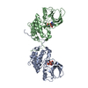
| |||||||||||||||||||||||||||||||||||||||
|---|---|---|---|---|---|---|---|---|---|---|---|---|---|---|---|---|---|---|---|---|---|---|---|---|---|---|---|---|---|---|---|---|---|---|---|---|---|---|---|---|
| 1 |
| |||||||||||||||||||||||||||||||||||||||
| 2 | 
| |||||||||||||||||||||||||||||||||||||||
| Unit cell |
| |||||||||||||||||||||||||||||||||||||||
| Noncrystallographic symmetry (NCS) | NCS domain:
NCS domain segments:
|
-Components
| #1: Protein | Mass: 36449.207 Da / Num. of mol.: 2 / Fragment: UNP residues 698-1019 Source method: isolated from a genetically manipulated source Source: (gene. exp.) Homo sapiens (human) / Gene: ERBB3, ErbB3/HER3, HER3 / Plasmid: pFASTBacHTa / Production host:   Spodoptera frugiperda (fall armyworm) / Strain (production host): Sf9 Spodoptera frugiperda (fall armyworm) / Strain (production host): Sf9References: UniProt: P21860, receptor protein-tyrosine kinase#2: Chemical |   Mass: 506.196 Da / Num. of mol.: 2 / Source method: obtained synthetically / Formula: C10H17N6O12P3 / Comment: AMP-PNP, energy-carrying molecule analogue*YM Mass: 506.196 Da / Num. of mol.: 2 / Source method: obtained synthetically / Formula: C10H17N6O12P3 / Comment: AMP-PNP, energy-carrying molecule analogue*YM#3: Chemical |   Mass: 24.305 Da / Num. of mol.: 2 / Source method: obtained synthetically / Formula: Mg Mass: 24.305 Da / Num. of mol.: 2 / Source method: obtained synthetically / Formula: Mg#4: Water | ChemComp-HOH / | Water Mass: 18.015 Da / Num. of mol.: 30 / Source method: isolated from a natural source / Formula: H2O Mass: 18.015 Da / Num. of mol.: 30 / Source method: isolated from a natural source / Formula: H2O |
|---|
-Experimental details
-Experiment
| Experiment | Method: X-RAY DIFFRACTION / Number of used crystals: 1 |
|---|
- Sample preparation
| Crystal | Density Matthews: 2.45 Å3/Da / Density % sol: 49.75 % |
|---|---|
| Crystal grow | Temperature: 293 K / Method: vapor diffusion, hanging drop / pH: 8.5 Details: 5mM AMP-PNP, 2mM MgCl2, 1mM DTT, 1mM TCEP, 0.1M Tris pH8.5, 0.2M lithium sulfate, 25% w/v PEG 3350 supplemented with 0.2% w/v 1,2- Diaminocyclohexane sulfate, 0.2% w/v Diloxanite Furoate, 0. ...Details: 5mM AMP-PNP, 2mM MgCl2, 1mM DTT, 1mM TCEP, 0.1M Tris pH8.5, 0.2M lithium sulfate, 25% w/v PEG 3350 supplemented with 0.2% w/v 1,2- Diaminocyclohexane sulfate, 0.2% w/v Diloxanite Furoate, 0.2% w/v Fumaric Acid, 0.2% w/v Spermine, 0.2% w/v Sulfaguanidine, 0.02M Hepes sodium pH 6.8, VAPOR DIFFUSION, HANGING DROP, temperature 293K |
-Data collection
| Diffraction | Mean temperature: 298 K |
|---|---|
| Diffraction source | Source: SYNCHROTRON / Site: ALS  / Beamline: 8.2.1 / Wavelength: 1 Å / Beamline: 8.2.1 / Wavelength: 1 Å |
| Detector | Type: ADSC QUANTUM 315r / Detector: CCD / Date: Apr 5, 2009 / Details: KOHZU: Double Crystal Si(111) |
| Radiation | Monochromator: KOHZU Double crystal, Si(111) / Protocol: SINGLE WAVELENGTH / Monochromatic (M) / Laue (L): M / Scattering type: x-ray |
| Radiation wavelength | Wavelength: 1 Å / Relative weight: 1 |
| Reflection | Resolution: 2.8→50 Å / Num. all: 17822 / Num. obs: 17502 / % possible obs: 98.2 % / Observed criterion σ(F): 1 / Observed criterion σ(I): 1 / Redundancy: 3.6 % / Biso Wilson estimate: 62.94 Å2 / Rmerge(I) obs: 0.101 / Χ2: 1 / Net I/σ(I): 6.9 |
| Reflection shell | Resolution: 2.8→2.9 Å / Redundancy: 3.2 % / Rmerge(I) obs: 0.553 / Mean I/σ(I) obs: 1.7 / Num. unique all: 1618 / Χ2: 0.725 / % possible all: 93 |
-Phasing
| Phasing | Method: molecular replacement | |||||||||
|---|---|---|---|---|---|---|---|---|---|---|
| Phasing MR | Rfactor: 53.3 / Model details: Phaser MODE: MR_AUTO
|
- Processing
| Software |
| |||||||||||||||||||||||||||||||||||||||||||||||||
|---|---|---|---|---|---|---|---|---|---|---|---|---|---|---|---|---|---|---|---|---|---|---|---|---|---|---|---|---|---|---|---|---|---|---|---|---|---|---|---|---|---|---|---|---|---|---|---|---|---|---|
| Refinement | Method to determine structure: MOLECULAR REPLACEMENT Starting model: inactive EGFR kinase domain Resolution: 2.797→46.463 Å / Occupancy max: 1 / Occupancy min: 1 / SU ML: 0.46 / σ(F): 1.93 / Stereochemistry target values: ML
| |||||||||||||||||||||||||||||||||||||||||||||||||
| Solvent computation | Shrinkage radii: 0.9 Å / VDW probe radii: 1.11 Å / Solvent model: FLAT BULK SOLVENT MODEL / Bsol: 61.617 Å2 / ksol: 0.323 e/Å3 | |||||||||||||||||||||||||||||||||||||||||||||||||
| Displacement parameters | Biso max: 158.72 Å2 / Biso mean: 71.101 Å2 / Biso min: 20 Å2
| |||||||||||||||||||||||||||||||||||||||||||||||||
| Refinement step | Cycle: LAST / Resolution: 2.797→46.463 Å
| |||||||||||||||||||||||||||||||||||||||||||||||||
| Refine LS restraints |
| |||||||||||||||||||||||||||||||||||||||||||||||||
| Refine LS restraints NCS |
| |||||||||||||||||||||||||||||||||||||||||||||||||
| LS refinement shell | Refine-ID: X-RAY DIFFRACTION / Total num. of bins used: 6
|
