 Movie
Movie Controller

[English] 日本語
 Yorodumi
- PDB-3kcg: Crystal structure of the antithrombin-factor IXa-pentasaccharide ... -
+ Open data

- Basic information
| Entry | Database: PDB / ID: 3kcg | |||||||||
|---|---|---|---|---|---|---|---|---|---|---|
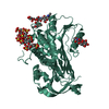
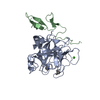
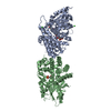
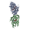
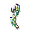
| Title | Crystal structure of the antithrombin-factor IXa-pentasaccharide complex | |||||||||
 Components Components |
| |||||||||
 Keywords Keywords |  HYDROLASE / HYDROLASE INHIBITOR / Michaelis complex / Blood coagulation / Calcium / Disulfide bond / EGF-like domain / Glycoprotein / Hemophilia / Pharmaceutical / Protease / Secreted / Serine protease / Sulfation / Zymogen / Heparin-binding / Protease inhibitor / Serine protease inhibitor / Thrombophilia HYDROLASE / HYDROLASE INHIBITOR / Michaelis complex / Blood coagulation / Calcium / Disulfide bond / EGF-like domain / Glycoprotein / Hemophilia / Pharmaceutical / Protease / Secreted / Serine protease / Sulfation / Zymogen / Heparin-binding / Protease inhibitor / Serine protease inhibitor / Thrombophilia | |||||||||
| Function / homology |  Function and homology information Function and homology informationDefective F9 secretion / Defective gamma-carboxylation of F9 / coagulation factor IXa / Defective F9 activation / Defective factor IX causes thrombophilia / Defective cofactor function of FVIIIa variant / Defective F9 variant does not activate FX / zymogen activation / Extrinsic Pathway of Fibrin Clot Formation / Protein hydroxylation ...Defective F9 secretion / Defective gamma-carboxylation of F9 / coagulation factor IXa / Defective F9 activation / Defective factor IX causes thrombophilia / Defective cofactor function of FVIIIa variant / Defective F9 variant does not activate FX / zymogen activation / Extrinsic Pathway of Fibrin Clot Formation / Protein hydroxylation / regulation of blood coagulation / Gamma-carboxylation of protein precursors / Transport of gamma-carboxylated protein precursors from the endoplasmic reticulum to the Golgi apparatus / Common Pathway of Fibrin Clot Formation / Removal of aminoterminal propeptides from gamma-carboxylated proteins / Intrinsic Pathway of Fibrin Clot Formation / Post-translational protein phosphorylation / serine-type endopeptidase inhibitor activity / Golgi lumen / Regulation of Insulin-like Growth Factor (IGF) transport and uptake by Insulin-like Growth Factor Binding Proteins (IGFBPs) / blood coagulation / heparin binding / collagen-containing extracellular matrix / endopeptidase activity / protease binding / blood microparticle / endoplasmic reticulum lumen / serine-type endopeptidase activity / calcium ion binding / proteolysis / extracellular space / extracellular exosome / extracellular region / identical protein binding / metal ion binding / plasma membraneSimilarity search - Function | |||||||||
| Biological species |  Homo sapiens (human) Homo sapiens (human) | |||||||||
| Method | X-RAY DIFFRACTION / SYNCHROTRON / MOLECULAR REPLACEMENT / Resolution: 1.7 Å | |||||||||
 Authors Authors | Huntington, J.A. / Johnson, D.J.D. | |||||||||
 Citation Citation | Journal: Proc.Natl.Acad.Sci.USA / Year: 2010 Title: Molecular basis of factor IXa recognition by heparin-activated antithrombin revealed by a 1.7-A structure of the ternary complex. Authors: Johnson, D.J. / Langdown, J. / Huntington, J.A. | |||||||||
| History |
|
- Structure visualization
| Structure viewer | Molecule: MolmilJmol/JSmol |
|---|
- Downloads & links
-Download
| PDBx/mmCIF format | 3kcg.cif.gz | 186.1 KB | Display | PDBx/mmCIF format |
|---|---|---|---|---|
| PDB format | pdb3kcg.ent.gz | 141.6 KB | Display | PDB format |
| PDBx/mmJSON format | 3kcg.json.gz | Tree view | PDBx/mmJSON format | |
| Others |  Other downloads Other downloads |
-Validation report
| Arichive directory | https://data.pdbj.org/pub/pdb/validation_reports/kc/3kcgftp://data.pdbj.org/pub/pdb/validation_reports/kc/3kcg | HTTPS FTP |
|---|
-Related structure data


-Links
 PDBj
PDBj


- Assembly
| Deposited unit | 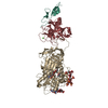
| ||||||||
|---|---|---|---|---|---|---|---|---|---|
| 1 |
| ||||||||
| Unit cell |
|
-Components
-Coagulation factor IXa ... , 2 types, 2 molecules LH
| #1: Protein | Factor IX / factor IX / Christmas factor / Plasma thromboplastin component / PTC Mass: 6470.354 Da / Num. of mol.: 1 / Fragment: EGF2 Source method: isolated from a genetically manipulated source Source: (gene. exp.) Homo sapiens (human) / Gene: F9 / Production host:  Escherichia coli (E. coli) / References: UniProt: P00740, coagulation factor IXa Escherichia coli (E. coli) / References: UniProt: P00740, coagulation factor IXa |
|---|---|
| #2: Protein | Factor IX / factor IX / Christmas factor / Plasma thromboplastin component / PTC Mass: 26174.818 Da / Num. of mol.: 1 / Mutation: S195A Source method: isolated from a genetically manipulated source Source: (gene. exp.) Homo sapiens (human) / Gene: F9 / Production host: Escherichia coli (E. coli) / References: UniProt: P00740, coagulation factor IXa |
-Protein , 1 types, 1 molecules I
| #3: Protein | / ATIII Mass: 49085.016 Da / Num. of mol.: 1 / Mutation: S137A Source method: isolated from a genetically manipulated source Source: (gene. exp.) Homo sapiens (human) / Gene: SERPINC1, AT3, PRO0309 / Cell (production host): BHK / References: UniProt: P01008 |
|---|
-Sugars , 3 types, 4 molecules
| #4: Polysaccharide | / Mass: 367.349 Da / Num. of mol.: 2 Source method: isolated from a genetically manipulated source #5: Polysaccharide | alpha-D-mannopyranose-(1-4)-2-acetamido-2-deoxy-beta-D-glucopyranose-(1-4)-2-acetamido-2-deoxy-beta- ...alpha-D-mannopyranose-(1-4)-2-acetamido-2-deoxy-beta-D-glucopyranose-(1-4)-2-acetamido-2-deoxy-beta-D-glucopyranose | / Mass: 586.542 Da / Num. of mol.: 1Source method: isolated from a genetically manipulated source #6: Polysaccharide | 3,4-di-O-methyl-2,6-di-O-sulfo-alpha-D-glucopyranose-(1-4)-2,3-di-O-methyl-beta-D-glucopyranuronic ...3,4-di-O-methyl-2,6-di-O-sulfo-alpha-D-glucopyranose-(1-4)-2,3-di-O-methyl-beta-D-glucopyranuronic acid-(1-4)-2,3,6-tri-O-sulfo-alpha-D-glucopyranose-(1-4)-3-O-methyl-2-O-sulfo-alpha-L-idopyranuronic acid-(1-4)-methyl 2,3,6-tri-O-sulfo-alpha-D-glucopyranoside / heparin pentasaccharide |   , Oligosaccharide / Class: Anticoagulant / Mass: 1661.413 Da / Num. of mol.: 1 , Oligosaccharide / Class: Anticoagulant / Mass: 1661.413 Da / Num. of mol.: 1Source method: isolated from a genetically manipulated source Details: oligosaccharide / References: heparin pentasaccharide |
|---|
-Non-polymers , 3 types, 670 molecules 


| #7: Chemical | ChemComp-CA /  Mass: 40.078 Da / Num. of mol.: 1 / Source method: obtained synthetically / Formula: Ca Mass: 40.078 Da / Num. of mol.: 1 / Source method: obtained synthetically / Formula: Ca | ||
|---|---|---|---|
| #8: Chemical | 2-Methyl-2,4-pentanediol Mass: 118.174 Da / Num. of mol.: 2 / Source method: obtained synthetically / Formula: C6H14O2 / Comment: precipitant*YM Mass: 118.174 Da / Num. of mol.: 2 / Source method: obtained synthetically / Formula: C6H14O2 / Comment: precipitant*YM#9: Water | ChemComp-HOH / | WaterMass: 18.015 Da / Num. of mol.: 667 / Source method: isolated from a natural source / Formula: H2O |
-Experimental details
-Experiment
| Experiment | Method: X-RAY DIFFRACTION / Number of used crystals: 1 |
|---|
- Sample preparation
| Crystal | Density Matthews: 3.14 Å3/Da / Density % sol: 60.8 % |
|---|---|
| Crystal grow | Temperature: 295 K / Method: vapor diffusion, hanging drop / pH: 7.4 Details: 0.25M Ammonium sulfate, 19.5% PEG 3350, pH 7.4, VAPOR DIFFUSION, HANGING DROP, temperature 295K |
-Data collection
| Diffraction | Mean temperature: 100 K |
|---|---|
| Diffraction source | Source: SYNCHROTRON / Site: Diamond  / Beamline: I02 / Wavelength: 0.9796 Å / Beamline: I02 / Wavelength: 0.9796 Å |
| Detector | Type: ADSC QUANTUM 315 / Detector: CCD / Date: May 24, 2009 |
| Radiation | Monochromator: double crystal / Protocol: SINGLE WAVELENGTH / Monochromatic (M) / Laue (L): M / Scattering type: x-ray |
| Radiation wavelength | Wavelength: 0.9796 Å / Relative weight: 1 |
| Reflection | Resolution: 1.7→58.83 Å / Num. all: 113381 / Num. obs: 112588 / % possible obs: 99.3 % / Observed criterion σ(F): 0 / Observed criterion σ(I): 0 / Redundancy: 5.2 % / Biso Wilson estimate: 26.7 Å2 / Rmerge(I) obs: 0.069 / Rsym value: 0.069 / Net I/σ(I): 5.7 |
| Reflection shell | Resolution: 1.7→1.79 Å / Redundancy: 3.6 % / Rmerge(I) obs: 0.58 / Mean I/σ(I) obs: 2.2 / Num. unique all: 16392 / Rsym value: 0.58 / % possible all: 100 |
- Processing
| Software |
| ||||||||||||||||||||||||||||||||||||
|---|---|---|---|---|---|---|---|---|---|---|---|---|---|---|---|---|---|---|---|---|---|---|---|---|---|---|---|---|---|---|---|---|---|---|---|---|---|
| Refinement | Method to determine structure: MOLECULAR REPLACEMENT Starting model: 1E03, 1RFN Resolution: 1.7→38.05 Å / Rfactor Rfree error: 0.003 / Data cutoff high absF: 2168682.34 / Data cutoff low absF: 0 / Isotropic thermal model: RESTRAINED / Cross valid method: THROUGHOUT / σ(F): 0 / Stereochemistry target values: Engh & Huber
| ||||||||||||||||||||||||||||||||||||
| Solvent computation | Solvent model: FLAT MODEL / Bsol: 52.4519 Å2 / ksol: 0.35 e/Å3 | ||||||||||||||||||||||||||||||||||||
| Displacement parameters | Biso mean: 33.3 Å2
| ||||||||||||||||||||||||||||||||||||
| Refine analyze |
| ||||||||||||||||||||||||||||||||||||
| Refinement step | Cycle: LAST / Resolution: 1.7→38.05 Å
| ||||||||||||||||||||||||||||||||||||
| Refine LS restraints |
| ||||||||||||||||||||||||||||||||||||
| LS refinement shell | Resolution: 1.7→1.81 Å / Rfactor Rfree error: 0.011 / Total num. of bins used: 6
|
