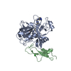
 Movie
Movie Controller


+ Open data

- Basic information
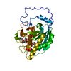
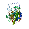
| Entry | Database: PDB / ID: 1rfn | ||||||
|---|---|---|---|---|---|---|---|
| Title | HUMAN COAGULATION FACTOR IXA IN COMPLEX WITH P-AMINO BENZAMIDINE | ||||||
 Components Components | (PROTEIN (COAGULATION FACTOR ...) x 2 | ||||||
 Keywords Keywords |  COAGULATION FACTOR / SERINE PROTEINASE / BLOOD COAGULATION COAGULATION FACTOR / SERINE PROTEINASE / BLOOD COAGULATION | ||||||
| Function / homology |  Function and homology information Function and homology informationDefective F9 secretion / Defective gamma-carboxylation of F9 / coagulation factor IXa / Defective F9 activation / Defective factor IX causes thrombophilia / Defective cofactor function of FVIIIa variant / Defective F9 variant does not activate FX / zymogen activation / Extrinsic Pathway of Fibrin Clot Formation / Protein hydroxylation ...Defective F9 secretion / Defective gamma-carboxylation of F9 / coagulation factor IXa / Defective F9 activation / Defective factor IX causes thrombophilia / Defective cofactor function of FVIIIa variant / Defective F9 variant does not activate FX / zymogen activation / Extrinsic Pathway of Fibrin Clot Formation / Protein hydroxylation / Gamma-carboxylation of protein precursors / Transport of gamma-carboxylated protein precursors from the endoplasmic reticulum to the Golgi apparatus / Removal of aminoterminal propeptides from gamma-carboxylated proteins / Intrinsic Pathway of Fibrin Clot Formation / Golgi lumen / blood coagulation / collagen-containing extracellular matrix / endopeptidase activity / endoplasmic reticulum lumen / serine-type endopeptidase activity / calcium ion binding / proteolysis / extracellular space / extracellular exosome / extracellular region / metal ion binding / plasma membraneSimilarity search - Function | ||||||
| Biological species |  Homo sapiens (human) Homo sapiens (human) | ||||||
| Method | X-RAY DIFFRACTION / MOLECULAR REPLACEMENT / Resolution: 2.8 Å | ||||||
 Authors Authors | Hopfner, K.-P. / Lang, A. / Karcher, A. / Sichler, K. / Kopetzki, E. / Brandstetter, H. / Huber, R. / Bode, W. / Engh, R.A. | ||||||
 Citation Citation | Journal: Structure Fold.Des. / Year: 1999 Title: Coagulation factor IXa: the relaxed conformation of Tyr99 blocks substrate binding. Authors: Hopfner, K.P. / Lang, A. / Karcher, A. / Sichler, K. / Kopetzki, E. / Brandstetter, H. / Huber, R. / Bode, W. / Engh, R.A. | ||||||
| History |
|
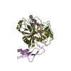
- Structure visualization
| Structure viewer | Molecule: MolmilJmol/JSmol |
|---|
- Downloads & links
-Download
| PDBx/mmCIF format | 1rfn.cif.gz | 69.6 KB | Display | PDBx/mmCIF format |
|---|---|---|---|---|
| PDB format | pdb1rfn.ent.gz | 51.8 KB | Display | PDB format |
| PDBx/mmJSON format | 1rfn.json.gz | Tree view | PDBx/mmJSON format | |
| Others |  Other downloads Other downloads |
-Validation report
| Arichive directory | https://data.pdbj.org/pub/pdb/validation_reports/rf/1rfnftp://data.pdbj.org/pub/pdb/validation_reports/rf/1rfn | HTTPS FTP |
|---|
-Related structure data
| Similar structure data |
|---|


-Links
 PDBj
PDBj


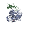
- Assembly
| Deposited unit | 
| ||||||||
|---|---|---|---|---|---|---|---|---|---|
| 1 |
| ||||||||
| Unit cell |
|
-Components
-PROTEIN (COAGULATION FACTOR ... , 2 types, 2 molecules AB
| #1: Protein | Mass: 26190.818 Da / Num. of mol.: 1 / Fragment: FRAGMENT EGF2-CATALYTIC DOMAIN Source method: isolated from a genetically manipulated source Source: (gene. exp.) Homo sapiens (human) / Production host:  Escherichia coli (E. coli) / Strain (production host): UT5600 / References: UniProt: P00740 Escherichia coli (E. coli) / Strain (production host): UT5600 / References: UniProt: P00740 |
|---|---|
| #2: Protein | Mass: 6256.135 Da / Num. of mol.: 1 / Fragment: FRAGMENT EGF2-CATALYTIC DOMAIN Source method: isolated from a genetically manipulated source Source: (gene. exp.) Homo sapiens (human) / Production host: Escherichia coli (E. coli) / Strain (production host): UT5600 / References: UniProt: P00740 |
-Non-polymers , 4 types, 19 molecules 

| #3: Chemical | ChemComp-CA /  Mass: 40.078 Da / Num. of mol.: 1 / Source method: obtained synthetically / Formula: Ca Mass: 40.078 Da / Num. of mol.: 1 / Source method: obtained synthetically / Formula: Ca |
|---|---|
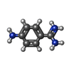
| #4: Chemical | ChemComp-PBZ /  Mass: 136.174 Da / Num. of mol.: 1 / Source method: obtained synthetically / Formula: C7H10N3 Mass: 136.174 Da / Num. of mol.: 1 / Source method: obtained synthetically / Formula: C7H10N3 |
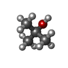
| #5: Chemical | ChemComp-TBU / Tert-Butyl alcohol Mass: 74.122 Da / Num. of mol.: 1 / Source method: obtained synthetically / Formula: C4H10O Mass: 74.122 Da / Num. of mol.: 1 / Source method: obtained synthetically / Formula: C4H10O |
| #6: Water | ChemComp-HOH / WaterMass: 18.015 Da / Num. of mol.: 16 / Source method: isolated from a natural source / Formula: H2O |
-Experimental details
-Experiment
| Experiment | Method: X-RAY DIFFRACTION |
|---|
- Sample preparation
| Crystal | Density Matthews: 2.79 Å3/Da / Density % sol: 55.84 % | ||||||||||||||||||||||||||||||||||||
|---|---|---|---|---|---|---|---|---|---|---|---|---|---|---|---|---|---|---|---|---|---|---|---|---|---|---|---|---|---|---|---|---|---|---|---|---|---|
| Crystal grow | pH: 8 / Details: pH 8.00 | ||||||||||||||||||||||||||||||||||||
| Crystal grow | *PLUS Temperature: 4 ℃ / Method: vapor diffusion, sitting drop | ||||||||||||||||||||||||||||||||||||
| Components of the solutions | *PLUS
|
-Data collection
| Diffraction | Mean temperature: 280 K |
|---|---|
| Diffraction source | Wavelength: 1.5418 |
| Radiation | Protocol: SINGLE WAVELENGTH / Monochromatic (M) / Laue (L): M / Scattering type: x-ray |
| Radiation wavelength | Wavelength: 1.5418 Å / Relative weight: 1 |
| Reflection | Resolution: 2.8→20 Å / Num. obs: 16348 / % possible obs: 92.7 % / Redundancy: 2.2 % / Rmerge(I) obs: 0.077 |
| Reflection | *PLUS Highest resolution: 2.8 Å / Lowest resolution: 20 Å / % possible obs: 92.7 % / Redundancy: 2.2 % |
| Reflection shell | *PLUS Highest resolution: 2.8 Å / Lowest resolution: 2.93 Å / % possible obs: 82 % / Rmerge(I) obs: 0.316 |
- Processing
| Software | Name: X-PLOR / Version: 3.8 / Classification: refinement | ||||||||||||||||||||||||||||||||||||||||||||||||||||||||||||
|---|---|---|---|---|---|---|---|---|---|---|---|---|---|---|---|---|---|---|---|---|---|---|---|---|---|---|---|---|---|---|---|---|---|---|---|---|---|---|---|---|---|---|---|---|---|---|---|---|---|---|---|---|---|---|---|---|---|---|---|---|---|
| Refinement | Method to determine structure: MOLECULAR REPLACEMENT / Resolution: 2.8→25 Å / Cross valid method: THROUGHOUT / σ(F): 0
| ||||||||||||||||||||||||||||||||||||||||||||||||||||||||||||
| Refinement step | Cycle: LAST / Resolution: 2.8→25 Å
| ||||||||||||||||||||||||||||||||||||||||||||||||||||||||||||
| Refine LS restraints |
| ||||||||||||||||||||||||||||||||||||||||||||||||||||||||||||
| Software | *PLUS Version: 3.8 / Classification: refinement | ||||||||||||||||||||||||||||||||||||||||||||||||||||||||||||
| Refinement | *PLUS Highest resolution: 2.8 Å / Lowest resolution: 25 Å / Rfactor obs: 0.216 | ||||||||||||||||||||||||||||||||||||||||||||||||||||||||||||
| Solvent computation | *PLUS | ||||||||||||||||||||||||||||||||||||||||||||||||||||||||||||
| Displacement parameters | *PLUS |
