 Movie
Movie Controller

[English] 日本語
 Yorodumi
- PDB-3k9t: Crystal structure of putative peptidase (NP_348812.1) from CLOSTR... -
+ Open data

- Basic information
| Entry | Database: PDB / ID: 3k9t | ||||||
|---|---|---|---|---|---|---|---|
| Title | Crystal structure of putative peptidase (NP_348812.1) from CLOSTRIDIUM ACETOBUTYLICUM at 2.37 A resolution | ||||||
 Components Components | putative peptidase Protease Protease | ||||||
 Keywords Keywords | HYDROLASE / putative peptidase / Structural Genomics / Joint Center for Structural Genomics / JCSG / Protein Structure Initiative / PSI-2 / Aminopeptidase | ||||||
| Function / homology |  Function and homology information Function and homology information | ||||||
| Biological species |  Clostridium acetobutylicum (bacteria) Clostridium acetobutylicum (bacteria) | ||||||
| Method | X-RAY DIFFRACTION / SYNCHROTRON / MAD / Resolution: 2.37 Å | ||||||
 Authors Authors | Joint Center for Structural Genomics (JCSG) | ||||||
 Citation Citation | Journal: To be published Title: Crystal structure of putative peptidase (NP_348812.1) from CLOSTRIDIUM ACETOBUTYLICUM at 2.37 A resolution Authors: Joint Center for Structural Genomics (JCSG) | ||||||
| History |
|
- Structure visualization
| Structure viewer | Molecule: MolmilJmol/JSmol |
|---|
- Downloads & links
-Download
| PDBx/mmCIF format | 3k9t.cif.gz | 103.8 KB | Display | PDBx/mmCIF format |
|---|---|---|---|---|
| PDB format | pdb3k9t.ent.gz | 82.1 KB | Display | PDB format |
| PDBx/mmJSON format | 3k9t.json.gz | Tree view | PDBx/mmJSON format | |
| Others |  Other downloads Other downloads |
-Validation report
| Arichive directory | https://data.pdbj.org/pub/pdb/validation_reports/k9/3k9tftp://data.pdbj.org/pub/pdb/validation_reports/k9/3k9t | HTTPS FTP |
|---|
-Related structure data
| Similar structure data | |
|---|---|
| Other databases |


-Links
 PDBj
PDBj


- Assembly
| Deposited unit | 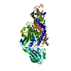
| ||||||||
|---|---|---|---|---|---|---|---|---|---|
| 1 | 
| ||||||||
| Unit cell |
| ||||||||
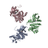
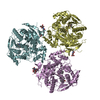
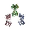
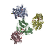
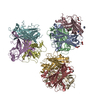
| Details | CRYSTAL PACKING ANALYSIS AND SIZE-EXCLUSION CHROMATOGRAPHY COUPLED WITH STATIC LIGHT SCATTERING SUPPORT THE ASSIGNMENT OF A TRIMER AS THE SIGNIFICANT OLIGOMERIC FORM IN SOLUTION. |
-Components
-Protein , 1 types, 1 molecules A
| #1: Protein | Protease / Protein containing aminopeptidase domain Mass: 50586.395 Da / Num. of mol.: 1 / Mutation: P309S Source method: isolated from a genetically manipulated source Source: (gene. exp.) Clostridium acetobutylicum (bacteria) / Gene: CA_C2195 / Plasmid: SpeedET / Production host: Escherichia Coli (E. coli) / Strain (production host): HK100 / References: UniProt: Q97H19 |
|---|
-Non-polymers , 5 types, 229 molecules 


| #2: Chemical | ChemComp-ZN /  Mass: 65.409 Da / Num. of mol.: 1 / Source method: obtained synthetically / Formula: Zn Mass: 65.409 Da / Num. of mol.: 1 / Source method: obtained synthetically / Formula: Zn | ||||||
|---|---|---|---|---|---|---|---|
| #3: Chemical | Chloride Mass: 35.453 Da / Num. of mol.: 2 / Source method: obtained synthetically / Formula: Cl Mass: 35.453 Da / Num. of mol.: 2 / Source method: obtained synthetically / Formula: Cl#4: Chemical | ChemComp-IMD / | Imidazole Mass: 69.085 Da / Num. of mol.: 1 / Source method: obtained synthetically / Formula: C3H5N2 Mass: 69.085 Da / Num. of mol.: 1 / Source method: obtained synthetically / Formula: C3H5N2#5: Chemical | ChemComp-MRD / ( 2-Methyl-2,4-pentanediol Mass: 118.174 Da / Num. of mol.: 4 / Source method: obtained synthetically / Formula: C6H14O2 / Comment: precipitant*YM Mass: 118.174 Da / Num. of mol.: 4 / Source method: obtained synthetically / Formula: C6H14O2 / Comment: precipitant*YM#6: Water | ChemComp-HOH / | WaterMass: 18.015 Da / Num. of mol.: 221 / Source method: isolated from a natural source / Formula: H2O |
-Details
| Sequence details | THIS CONSTRUCT WAS EXPRESSED WITH A PURIFICATION TAG MGSDKIHHHHHHENLYFQG. THE TAG WAS REMOVED WITH ...THIS CONSTRUCT WAS EXPRESSED WITH A PURIFICATI |
|---|
-Experimental details
-Experiment
| Experiment | Method: X-RAY DIFFRACTION / Number of used crystals: 1 |
|---|
- Sample preparation
| Crystal | Density Matthews: 3.79 Å3/Da / Density % sol: 67.52 % |
|---|---|
| Crystal grow | Temperature: 293 K / Method: vapor diffusion, sitting drop / pH: 8 Details: 0.2000M MgCl2, 40.0000% MPD, 0.1M Imidazole pH 8.0, Additive: 0.006M Zinc Chloride, NANODROP', VAPOR DIFFUSION, SITTING DROP, temperature 293K |
-Data collection
| Diffraction | Mean temperature: 100 K | |||||||||||||||||||||||||||||||||||||||||||||||||||||||||||||||||||||||||||||||||||||||||||||||||||||||||||||||||||||||||||||||||||||||||||||||||||
|---|---|---|---|---|---|---|---|---|---|---|---|---|---|---|---|---|---|---|---|---|---|---|---|---|---|---|---|---|---|---|---|---|---|---|---|---|---|---|---|---|---|---|---|---|---|---|---|---|---|---|---|---|---|---|---|---|---|---|---|---|---|---|---|---|---|---|---|---|---|---|---|---|---|---|---|---|---|---|---|---|---|---|---|---|---|---|---|---|---|---|---|---|---|---|---|---|---|---|---|---|---|---|---|---|---|---|---|---|---|---|---|---|---|---|---|---|---|---|---|---|---|---|---|---|---|---|---|---|---|---|---|---|---|---|---|---|---|---|---|---|---|---|---|---|---|---|---|---|
| Diffraction source | Source: SYNCHROTRON / Site: SSRL  / Beamline: BL9-2 / Wavelength: 0.91837,0.97911 / Beamline: BL9-2 / Wavelength: 0.91837,0.97911 | |||||||||||||||||||||||||||||||||||||||||||||||||||||||||||||||||||||||||||||||||||||||||||||||||||||||||||||||||||||||||||||||||||||||||||||||||||
| Detector | Type: MARMOSAIC 325 mm CCD / Detector: CCD / Date: Jul 31, 2009 / Details: Flat collimating mirror, toroid focusing mirror | |||||||||||||||||||||||||||||||||||||||||||||||||||||||||||||||||||||||||||||||||||||||||||||||||||||||||||||||||||||||||||||||||||||||||||||||||||
| Radiation | Monochromator: Double crystal monochromator / Protocol: MAD / Monochromatic (M) / Laue (L): M / Scattering type: x-ray | |||||||||||||||||||||||||||||||||||||||||||||||||||||||||||||||||||||||||||||||||||||||||||||||||||||||||||||||||||||||||||||||||||||||||||||||||||
| Radiation wavelength |
| |||||||||||||||||||||||||||||||||||||||||||||||||||||||||||||||||||||||||||||||||||||||||||||||||||||||||||||||||||||||||||||||||||||||||||||||||||
| Reflection | Resolution: 2.37→29.062 Å / Num. obs: 31178 / % possible obs: 99.9 % / Redundancy: 5.5 % / Biso Wilson estimate: 38.179 Å2 / Rmerge(I) obs: 0.189 / Rsym value: 0.189 / Net I/σ(I): 9 | |||||||||||||||||||||||||||||||||||||||||||||||||||||||||||||||||||||||||||||||||||||||||||||||||||||||||||||||||||||||||||||||||||||||||||||||||||
| Reflection shell | Rmerge(I) obs: 0.01 / Diffraction-ID: 1
|
-Phasing
| Phasing | Method: MAD |
|---|
- Processing
| Software |
| |||||||||||||||||||||||||||||||||||||||||||||||||||||||||||||||||||||||||||||||||||||
|---|---|---|---|---|---|---|---|---|---|---|---|---|---|---|---|---|---|---|---|---|---|---|---|---|---|---|---|---|---|---|---|---|---|---|---|---|---|---|---|---|---|---|---|---|---|---|---|---|---|---|---|---|---|---|---|---|---|---|---|---|---|---|---|---|---|---|---|---|---|---|---|---|---|---|---|---|---|---|---|---|---|---|---|---|---|---|
| Refinement | Method to determine structure: MAD / Resolution: 2.37→29.062 Å / Cor.coef. Fo:Fc: 0.956 / Cor.coef. Fo:Fc free: 0.935 / Occupancy max: 1 / Occupancy min: 0.4 / SU B: 11.776 / SU ML: 0.122 / TLS residual ADP flag: LIKELY RESIDUAL / Cross valid method: THROUGHOUT / σ(F): 0 / ESU R: 0.201 / ESU R Free: 0.18 Stereochemistry target values: MAXIMUM LIKELIHOOD WITH PHASES Details: 1.HYDROGENS HAVE BEEN ADDED IN THE RIDING POSITIONS. 2.ATOM RECORDS CONTAIN RESIDUAL B FACTORS ONLY. 3.A MET-INHIBITION PROTOCOL WAS USED FOR SELENOMETHIONINE INCORPORATION DURING PROTEIN ...Details: 1.HYDROGENS HAVE BEEN ADDED IN THE RIDING POSITIONS. 2.ATOM RECORDS CONTAIN RESIDUAL B FACTORS ONLY. 3.A MET-INHIBITION PROTOCOL WAS USED FOR SELENOMETHIONINE INCORPORATION DURING PROTEIN EXPRESSION. THE OCCUPANCY OF THE SE ATOMS IN THE MSE RESIDUES WAS REDUCED TO 0.75 FOR THE REDUCED SCATTERING POWER DUE TO PARTIAL S-MET INCORPORATION. 4.ZINC (ZN) HAS BEEN MODELED AT THE PUTATIVE ACTIVE SITE BASED ON ITS PRESENCE AS A CO-CRYSTALLIZATION COMPOUND AND ANOMALOUS DIFFERENCE FOURIER MAP. 5.CHLORIDE (CL), IMIDAZOLE (IMD) AND (4R)-2-METHYLPENTANE-2,4-DIOL (MRD) FROM THE CRYSTALLIZATION SOLUTION HAVE BEEN MODELED IN THE SOLVENT STRUCTURE. 6.THE RAMACHANDRAN OUTLIER AT RESIDUE PRO190 IS SUPPORTED BY ELECTRON DENSITY.
| |||||||||||||||||||||||||||||||||||||||||||||||||||||||||||||||||||||||||||||||||||||
| Solvent computation | Ion probe radii: 0.8 Å / Shrinkage radii: 0.8 Å / VDW probe radii: 1.2 Å / Solvent model: MASK | |||||||||||||||||||||||||||||||||||||||||||||||||||||||||||||||||||||||||||||||||||||
| Displacement parameters | Biso max: 384.4 Å2 / Biso mean: 29.498 Å2 / Biso min: 2 Å2
| |||||||||||||||||||||||||||||||||||||||||||||||||||||||||||||||||||||||||||||||||||||
| Refinement step | Cycle: LAST / Resolution: 2.37→29.062 Å
| |||||||||||||||||||||||||||||||||||||||||||||||||||||||||||||||||||||||||||||||||||||
| Refine LS restraints |
| |||||||||||||||||||||||||||||||||||||||||||||||||||||||||||||||||||||||||||||||||||||
| LS refinement shell | Resolution: 2.37→2.431 Å / Total num. of bins used: 20
| |||||||||||||||||||||||||||||||||||||||||||||||||||||||||||||||||||||||||||||||||||||
| Refinement TLS params. | Method: refined / Origin x: 32.2688 Å / Origin y: 1.6738 Å / Origin z: 64.0806 Å
|
