 Movie
Movie Controller


+ Open data

- Basic information
| Entry | Database: PDB / ID: 5d7v | ||||||
|---|---|---|---|---|---|---|---|
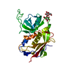
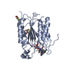
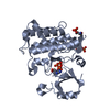
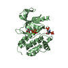
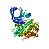
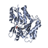
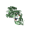
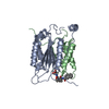
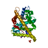
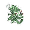
| Title | Crystal structure of PTK6 kinase domain | ||||||
 Components Components | Protein-tyrosine kinase 6 Tyrosine kinase Tyrosine kinase | ||||||
 Keywords Keywords | TRANSFERASE / KINASE / BRK / APO | ||||||
| Function / homology |  Function and homology information Function and homology informationPTK6 Activates STAT3 / negative regulation of protein tyrosine kinase activity / PTK6 Regulates Proteins Involved in RNA Processing / intestinal epithelial cell differentiation / ERBB2 signaling pathway / negative regulation of growth / PTK6 Expression / tyrosine phosphorylation of STAT protein / positive regulation of epidermal growth factor receptor signaling pathway / PTK6 promotes HIF1A stabilization ...PTK6 Activates STAT3 / negative regulation of protein tyrosine kinase activity / PTK6 Regulates Proteins Involved in RNA Processing / intestinal epithelial cell differentiation / ERBB2 signaling pathway / negative regulation of growth / PTK6 Expression / tyrosine phosphorylation of STAT protein / positive regulation of epidermal growth factor receptor signaling pathway / PTK6 promotes HIF1A stabilization / ERBB2 Activates PTK6 Signaling / PTK6 Down-Regulation / PTK6 Regulates Cell Cycle / PTK6 Regulates RHO GTPases, RAS GTPase and MAP kinases / positive regulation of cell cycle / cellular response to retinoic acid / positive regulation of tyrosine phosphorylation of STAT protein / ruffle / PTK6 Regulates RTKs and Their Effectors AKT1 and DOK1 / extrinsic component of cytoplasmic side of plasma membrane / positive regulation of DNA replication / non-specific protein-tyrosine kinase / non-membrane spanning protein tyrosine kinase activity / Cytoprotection by HMOX1 / SCF(Skp2)-mediated degradation of p27/p21 / positive regulation of neuron projection development / cell surface receptor protein tyrosine kinase signaling pathway / Cyclin D associated events in G1 / cell migration / protein tyrosine kinase activity / protein autophosphorylation / cell differentiation / nuclear body / protein phosphorylation / signaling receptor binding / innate immune response / nucleoplasm / ATP binding / identical protein binding / nucleus / plasma membrane / cytosol / cytoplasmSimilarity search - Function | ||||||
| Biological species |  Homo sapiens (human) Homo sapiens (human) | ||||||
| Method | X-RAY DIFFRACTION / MOLECULAR REPLACEMENT / Resolution: 2.33 Å | ||||||
| Model details | TRANSFERASE | ||||||
 Authors Authors | Thakur, M.K. / Birudukota, S. / Swaminathan, S. / Tyagi, R. / Gosu, R. | ||||||
 Citation Citation | Journal: Biochem.Biophys.Res.Commun. / Year: 2016 Title: Crystal structure of the kinase domain of human protein tyrosine kinase 6 (PTK6) at 2.33 angstrom resolution Authors: Thakur, M.K. / Kumar, A. / Birudukota, S. / Swaminathan, S. / Tyagi, R. / Gosu, R. | ||||||
| History |
|
- Structure visualization
| Structure viewer | Molecule: MolmilJmol/JSmol |
|---|
- Downloads & links
-Download
| PDBx/mmCIF format | 5d7v.cif.gz | 229.8 KB | Display | PDBx/mmCIF format |
|---|---|---|---|---|
| PDB format | pdb5d7v.ent.gz | 184.7 KB | Display | PDB format |
| PDBx/mmJSON format | 5d7v.json.gz | Tree view | PDBx/mmJSON format | |
| Others |  Other downloads Other downloads |
-Validation report
| Arichive directory | https://data.pdbj.org/pub/pdb/validation_reports/d7/5d7vftp://data.pdbj.org/pub/pdb/validation_reports/d7/5d7v | HTTPS FTP |
|---|
-Related structure data
| Related structure data | 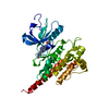 2f4jS S: Starting model for refinement |
|---|---|
| Similar structure data |
-Links
 PDBj
PDBj


- Assembly
| Deposited unit | 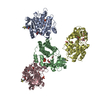
| ||||||||
|---|---|---|---|---|---|---|---|---|---|
| 1 | 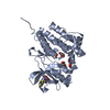
| ||||||||
| 2 | 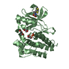
| ||||||||
| 3 | 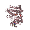
| ||||||||
| 4 | 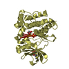
| ||||||||
| Unit cell |
|
-Components
| #1: Protein | Tyrosine kinase / Breast tumor kinase / Tyrosine-protein kinase BRK Mass: 31140.910 Da / Num. of mol.: 4 / Fragment: PTK6_KINASE DOMAIN, UNP residues 185-446 / Mutation: C433T Source method: isolated from a genetically manipulated source Source: (gene. exp.) Homo sapiens (human) / Gene: PTK6 / Plasmid: pFastBac1 / Cell line (production host): SF9 / Production host:   Spodoptera frugiperda (fall armyworm) Spodoptera frugiperda (fall armyworm)References: UniProt: Q13882, non-specific protein-tyrosine kinase#2: Chemical | ChemComp-PO4 / Phosphate  Mass: 94.971 Da / Num. of mol.: 4 / Source method: obtained synthetically / Formula: PO4 Mass: 94.971 Da / Num. of mol.: 4 / Source method: obtained synthetically / Formula: PO4#3: Chemical | ChemComp-GOL / Glycerol  Mass: 92.094 Da / Num. of mol.: 12 / Source method: obtained synthetically / Formula: C3H8O3 Mass: 92.094 Da / Num. of mol.: 12 / Source method: obtained synthetically / Formula: C3H8O3#4: Water | ChemComp-HOH / | Water Mass: 18.015 Da / Num. of mol.: 366 / Source method: isolated from a natural source / Formula: H2O Mass: 18.015 Da / Num. of mol.: 366 / Source method: isolated from a natural source / Formula: H2O |
|---|
-Experimental details
-Experiment
| Experiment | Method: X-RAY DIFFRACTION / Number of used crystals: 1 |
|---|
- Sample preparation
| Crystal | Density Matthews: 2.48 Å3/Da / Density % sol: 50.39 % |
|---|---|
| Crystal grow | Temperature: 298 K / Method: vapor diffusion, sitting drop Details: 0.23M Diammonium Phosphate, 18% PEG 3350, 1M Lithium chloride |
-Data collection
| Diffraction | Mean temperature: 100 K |
|---|---|
| Diffraction source | Source: ROTATING ANODE / Type: RIGAKU MICROMAX-007 HF / Wavelength: 1.54789 Å |
| Detector | Type: MAR scanner 345 mm plate / Detector: IMAGE PLATE / Date: Mar 3, 2015 / Details: VariMax HR |
| Radiation | Protocol: SINGLE WAVELENGTH / Monochromatic (M) / Laue (L): M / Scattering type: x-ray |
| Radiation wavelength | Wavelength: 1.54789 Å / Relative weight: 1 |
| Reflection | Resolution: 2.33→50 Å / Num. obs: 50742 / % possible obs: 96.3 % / Redundancy: 2.1 % / Rmerge(I) obs: 0.113 / Net I/σ(I): 7.3 |
| Reflection shell | Resolution: 2.33→2.41 Å / Redundancy: 2.1 % / Rmerge(I) obs: 0.443 / Mean I/σ(I) obs: 1.9 / % possible all: 94.2 |
- Processing
| Software |
| ||||||||||||||||||||||||||||||||||||||||||||||||||||||||||||||||||||||||||||||||||||||||||
|---|---|---|---|---|---|---|---|---|---|---|---|---|---|---|---|---|---|---|---|---|---|---|---|---|---|---|---|---|---|---|---|---|---|---|---|---|---|---|---|---|---|---|---|---|---|---|---|---|---|---|---|---|---|---|---|---|---|---|---|---|---|---|---|---|---|---|---|---|---|---|---|---|---|---|---|---|---|---|---|---|---|---|---|---|---|---|---|---|---|---|---|
| Refinement | Method to determine structure: MOLECULAR REPLACEMENT Starting model: 2F4J Resolution: 2.33→50 Å / Cor.coef. Fo:Fc: 0.925 / Cor.coef. Fo:Fc free: 0.88 / SU B: 9.7 / SU ML: 0.232 / Cross valid method: THROUGHOUT / σ(F): 0 / ESU R: 0.508 / ESU R Free: 0.301 / Stereochemistry target values: MAXIMUM LIKELIHOOD / Details: HYDROGENS HAVE BEEN ADDED IN THE RIDING POSITIONS
| ||||||||||||||||||||||||||||||||||||||||||||||||||||||||||||||||||||||||||||||||||||||||||
| Solvent computation | Ion probe radii: 0.8 Å / Shrinkage radii: 0.8 Å / VDW probe radii: 1.2 Å / Solvent model: MASK | ||||||||||||||||||||||||||||||||||||||||||||||||||||||||||||||||||||||||||||||||||||||||||
| Displacement parameters | Biso max: 72.01 Å2 / Biso mean: 26.633 Å2 / Biso min: 11.27 Å2
| ||||||||||||||||||||||||||||||||||||||||||||||||||||||||||||||||||||||||||||||||||||||||||
| Refinement step | Cycle: final / Resolution: 2.33→50 Å
| ||||||||||||||||||||||||||||||||||||||||||||||||||||||||||||||||||||||||||||||||||||||||||
| Refine LS restraints |
| ||||||||||||||||||||||||||||||||||||||||||||||||||||||||||||||||||||||||||||||||||||||||||
| LS refinement shell | Resolution: 2.334→2.394 Å / Total num. of bins used: 20
|
