 Movie
Movie Controller

[English] 日本語
 Yorodumi
- PDB-2g04: Crystal structure of fatty acid-CoA racemase from Mycobacterium t... -
+ Open data

- Basic information
| Entry | Database: PDB / ID: 2g04 | ||||||
|---|---|---|---|---|---|---|---|
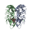
| Title | Crystal structure of fatty acid-CoA racemase from Mycobacterium tuberculosis H37Rv | ||||||
 Components Components | PROBABLE FATTY-ACID-CoA RACEMASE FAR | ||||||
 Keywords Keywords |  ISOMERASE ISOMERASE | ||||||
| Function / homology |  Function and homology information Function and homology information | ||||||
| Biological species |   Mycobacterium tuberculosis (bacteria) Mycobacterium tuberculosis (bacteria) | ||||||
| Method | X-RAY DIFFRACTION / SYNCHROTRON / MOLECULAR REPLACEMENT / Resolution: 2.7 Å | ||||||
 Authors Authors | Lee, K.S. / Park, S.M. / Rhee, K.H. / Bang, W.G. / Hwang, K.Y. / Chi, Y.M. | ||||||
 Citation Citation | Journal: Proteins / Year: 2006 Title: Crystal structure of fatty acid-CoA racemase from Mycobacterium tuberculosis H37Rv Authors: Lee, K.S. / Park, S.M. / Rhee, K.H. / Bang, W.G. / Hwang, K.Y. / Chi, Y.M. | ||||||
| History |
|
- Structure visualization
| Structure viewer | Molecule: MolmilJmol/JSmol |
|---|
- Downloads & links
-Download
| PDBx/mmCIF format | 2g04.cif.gz | 393.2 KB | Display | PDBx/mmCIF format |
|---|---|---|---|---|
| PDB format | pdb2g04.ent.gz | 324.4 KB | Display | PDB format |
| PDBx/mmJSON format | 2g04.json.gz | Tree view | PDBx/mmJSON format | |
| Others |  Other downloads Other downloads |
-Validation report
| Arichive directory | https://data.pdbj.org/pub/pdb/validation_reports/g0/2g04ftp://data.pdbj.org/pub/pdb/validation_reports/g0/2g04 | HTTPS FTP |
|---|
-Related structure data
| Related structure data |  1x74S S: Starting model for refinement |
|---|---|
| Similar structure data |


-Links
 PDBj
PDBj- Assembly
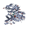
| Deposited unit | 
| ||||||||
|---|---|---|---|---|---|---|---|---|---|
| 1 | 
| ||||||||
| 2 | 
| ||||||||
| 3 | 
| ||||||||
| Unit cell |
|
-Components
| #1: Protein | Mass: 38094.441 Da / Num. of mol.: 6 Source method: isolated from a genetically manipulated source Source: (gene. exp.) Mycobacterium tuberculosis (bacteria) / Strain: H37Rv / Gene: far / Plasmid: pET-28a / Species (production host): Escherichia coli / Production host: Escherichia coli BL21(DE3) (bacteria) / Strain (production host): BL21(DE3) / References: UniProt: O53867, alpha-methylacyl-CoA racemase#2: Water | ChemComp-HOH / | Water Mass: 18.015 Da / Num. of mol.: 472 / Source method: isolated from a natural source / Formula: H2O Mass: 18.015 Da / Num. of mol.: 472 / Source method: isolated from a natural source / Formula: H2O |
|---|
-Experimental details
-Experiment
| Experiment | Method: X-RAY DIFFRACTION / Number of used crystals: 1 |
|---|
- Sample preparation
| Crystal | Density Matthews: 2.24 Å3/Da / Density % sol: 45.13 % |
|---|---|
| Crystal grow | Temperature: 295 K / Method: vapor diffusion, hanging drop / pH: 8.5 Details: 20% PEG 4000, 0.2M Amm, chloride, 0.01M Ca, chloride, pH 8.5, VAPOR DIFFUSION, HANGING DROP, temperature 295K |
-Data collection
| Diffraction | Mean temperature: 100 K |
|---|---|
| Diffraction source | Source: SYNCHROTRON / Site: PAL/PLS  / Beamline: 6B / Wavelength: 1.12714 Å / Beamline: 6B / Wavelength: 1.12714 Å |
| Detector | Type: BRUKER PROTEUM 300 / Detector: CCD / Date: Dec 4, 2005 |
| Radiation | Monochromator: Double Crystal monochromator / Protocol: SINGLE WAVELENGTH / Monochromatic (M) / Laue (L): M / Scattering type: x-ray |
| Radiation wavelength | Wavelength: 1.12714 Å / Relative weight: 1 |
| Reflection | Resolution: 2.7→50 Å / Num. obs: 52589 / % possible obs: 95.8 % / Observed criterion σ(F): 0 / Observed criterion σ(I): 0 / Redundancy: 3.7 % / Biso Wilson estimate: 35 Å2 / Rmerge(I) obs: 0.08 / Rsym value: 0.08 / Net I/σ(I): 9.9 |
| Reflection shell | Resolution: 2.7→2.82 Å / Redundancy: 4 % / Mean I/σ(I) obs: 2 / Num. unique all: 5942 / Rsym value: 0.306 / % possible all: 86.2 |
- Processing
| Software |
| |||||||||||||||||||||||||
|---|---|---|---|---|---|---|---|---|---|---|---|---|---|---|---|---|---|---|---|---|---|---|---|---|---|---|
| Refinement | Method to determine structure: MOLECULAR REPLACEMENT Starting model: PDB ENTRY 1X74 Resolution: 2.7→30 Å / Cross valid method: THROUGHOUT / σ(F): 0 / σ(I): 0
| |||||||||||||||||||||||||
| Displacement parameters | Biso mean: 55.9 Å2
| |||||||||||||||||||||||||
| Refine analyze |
| |||||||||||||||||||||||||
| Refinement step | Cycle: LAST / Resolution: 2.7→30 Å
| |||||||||||||||||||||||||
| Refine LS restraints |
| |||||||||||||||||||||||||
| LS refinement shell | Resolution: 2.7→2.87 Å / Rfactor Rfree error: 0.006
|
