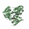
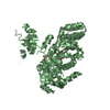
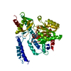
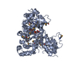
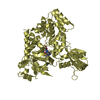
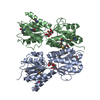
transcription regulator / STRUCTURAL GENOMICS / PROTEIN STRUCTURE INITIATIVE / NEW YORK STRUCTURAL GENOMIX RESEARCH CONSORTIUM / NYSGXRC / DNA-binding / Transcription / Transcription regulation / PSI-2 / New York SGX Research Center for Structural Genomics
Protocol: SINGLE WAVELENGTH / Scattering type: x-ray
Radiation wavelength
Wavelength: 0.9791 Å / Relative weight: 1
Reflection
Resolution: 2.7→50 Å / Num. obs: 48261 / % possible obs: 99.9 % / Redundancy: 6.4 % / Rmerge(I) obs: 0.096 / Net I/σ(I): 8
Reflection shell
Resolution: 2.7→2.75 Å / Redundancy: 4.9 % / % possible all: 99.5
-
Processing
Software
Name
Version
Classification
NB
DENZO
datareduction
SCALEPACK
datascaling
REFMAC
5.5.0089
refinement
PDB_EXTRACT
3.005
dataextraction
HKL-2000
datareduction
SHELXS
phasing
Refinement
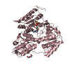
Method to determine structure: SAD / Resolution: 2.8→20 Å / Cor.coef. Fo:Fc: 0.943 / Cor.coef. Fo:Fc free: 0.911 / Occupancy max: 1 / Occupancy min: 0.4 / SU B: 21.107 / SU ML: 0.181 / TLS residual ADP flag: LIKELY RESIDUAL / Cross valid method: THROUGHOUT / ESU R: 0.123 / ESU R Free: 0.066 / Stereochemistry target values: MAXIMUM LIKELIHOOD Details: HYDROGENS HAVE BEEN ADDED IN THE RIDING POSITIONS. The maltose binding is only suggested based a) on the shape of electron density, b) the fact that other members of this group of proteins ...Details: HYDROGENS HAVE BEEN ADDED IN THE RIDING POSITIONS. The maltose binding is only suggested based a) on the shape of electron density, b) the fact that other members of this group of proteins bind maltose or lactose, c) suggested inhibitor makes multiple favorable contacts in the binding site. Lactose does not fit the density, but we can not exclude that some other similar sugar is bound. PLEASE REMOVE "complexed with maltose" from the title, and just add remark that maltose binding is SUGGESTED.
Rfactor
Num. reflection
% reflection
Selection details
Rfree
0.24388
1191
5.1 %
RANDOM
Rwork
0.20297
-
-
-
obs
0.20503
22103
99.59 %
-
Solvent computation
Ion probe radii: 0.8 Å / Shrinkage radii: 0.8 Å / VDW probe radii: 1.4 Å / Solvent model: BABINET MODEL WITH MASK
In the structure databanks used in Yorodumi, some data are registered as the other names, "COVID-19 virus" and "2019-nCoV". Here are the details of the virus and the list of structure data.
Jan 31, 2019. EMDB accession codes are about to change! (news from PDBe EMDB page)
EMDB accession codes are about to change! (news from PDBe EMDB page)
The allocation of 4 digits for EMDB accession codes will soon come to an end. Whilst these codes will remain in use, new EMDB accession codes will include an additional digit and will expand incrementally as the available range of codes is exhausted. The current 4-digit format prefixed with “EMD-” (i.e. EMD-XXXX) will advance to a 5-digit format (i.e. EMD-XXXXX), and so on. It is currently estimated that the 4-digit codes will be depleted around Spring 2019, at which point the 5-digit format will come into force.
The EM Navigator/Yorodumi systems omit the EMD- prefix.
Related info.:Q: What is EMD? / ID/Accession-code notation in Yorodumi/EM Navigator
Yorodumi is a browser for structure data from EMDB, PDB, SASBDB, etc.
This page is also the successor to EM Navigator detail page, and also detail information page/front-end page for Omokage search.
The word "yorodu" (or yorozu) is an old Japanese word meaning "ten thousand". "mi" (miru) is to see.
Related info.:EMDB / PDB / SASBDB / Comparison of 3 databanks / Yorodumi Search / Aug 31, 2016. New EM Navigator & Yorodumi / Yorodumi Papers / Jmol/JSmol / Function and homology information / Changes in new EM Navigator and Yorodumi
 Movie
Movie Controller

 Yorodumi
 Open data
 Basic information
 Components
 Transcriptional regulation
  Keywords
 Function and homology information
 Authors
 Citation
 Structure visualization
 Downloads & links
 Other downloads
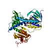

 PDBj
PDBj


 Assembly
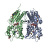


 Mass: 18.015 Da / Num. of mol.: 24 / Source method: isolated from a natural source / Formula: H2O
 Sample preparation
 / Beamline: X29A / Wavelength: 0.9791
  Processing