 Movie
Movie Controller


+ Open data

- Basic information
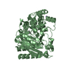
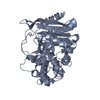
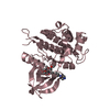
| Entry | Database: PDB / ID: 3k43 | ||||||
|---|---|---|---|---|---|---|---|
| Title | Crystal structure of sCD-MPR mutant E19Q/K137M pH 6.5 | ||||||
 Components Components | Cation-dependent mannose-6-phosphate receptor | ||||||
 Keywords Keywords | protein transport / sugar binding protein / transport / lysosome / mannose / receptor / sugar binding / Glycoprotein / Membrane / Phosphoprotein / Transmembrane | ||||||
| Function / homology |  Function and homology informationRetrograde transport at the Trans-Golgi-Network / Glycosphingolipid catabolism / Lysosome Vesicle Biogenesis / Cargo recognition for clathrin-mediated endocytosis / protein targeting to lysosome / Clathrin-mediated endocytosis / lysosomal transport / trans-Golgi network / endosome / lysosomal membrane ...Retrograde transport at the Trans-Golgi-Network / Glycosphingolipid catabolism / Lysosome Vesicle Biogenesis / Cargo recognition for clathrin-mediated endocytosis / protein targeting to lysosome / Clathrin-mediated endocytosis / lysosomal transport / trans-Golgi network / endosome / lysosomal membrane / protein domain specific binding / Golgi apparatus Function and homology informationRetrograde transport at the Trans-Golgi-Network / Glycosphingolipid catabolism / Lysosome Vesicle Biogenesis / Cargo recognition for clathrin-mediated endocytosis / protein targeting to lysosome / Clathrin-mediated endocytosis / lysosomal transport / trans-Golgi network / endosome / lysosomal membrane ...Retrograde transport at the Trans-Golgi-Network / Glycosphingolipid catabolism / Lysosome Vesicle Biogenesis / Cargo recognition for clathrin-mediated endocytosis / protein targeting to lysosome / Clathrin-mediated endocytosis / lysosomal transport / trans-Golgi network / endosome / lysosomal membrane / protein domain specific binding / Golgi apparatusSimilarity search - Function | ||||||
| Biological species |  Bos taurus (cattle) Bos taurus (cattle) | ||||||
| Method | X-RAY DIFFRACTION / difference fourier / Resolution: 2 Å | ||||||
 Authors Authors | Olson, L.J. / Sun, G. / Bohnsack, R.N. / Peterson, F.C. / Dahms, N.M. / Kim, J.J.P. | ||||||
 Citation Citation | Journal: Biochemistry / Year: 2010 Title: Intermonomer interactions are essential for lysosomal enzyme binding by the cation-dependent mannose 6-phosphate receptor. Authors: Olson, L.J. / Sun, G. / Bohnsack, R.N. / Peterson, F.C. / Dahms, N.M. / Kim, J.J. | ||||||
| History |
|
- Structure visualization
| Structure viewer | Molecule: MolmilJmol/JSmol |
|---|
- Downloads & links
-Download
| PDBx/mmCIF format | 3k43.cif.gz | 76.4 KB | Display | PDBx/mmCIF format |
|---|---|---|---|---|
| PDB format | pdb3k43.ent.gz | 56.2 KB | Display | PDB format |
| PDBx/mmJSON format | 3k43.json.gz | Tree view | PDBx/mmJSON format | |
| Others |  Other downloads Other downloads |
-Validation report
| Arichive directory | https://data.pdbj.org/pub/pdb/validation_reports/k4/3k43ftp://data.pdbj.org/pub/pdb/validation_reports/k4/3k43 | HTTPS FTP |
|---|
-Related structure data
| Related structure data | 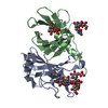 3k41C 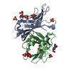 3k42SC S: Starting model for refinement C: citing same article ( |
|---|---|
| Similar structure data |


-Links
 PDBj
PDBj


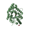
- Assembly
| Deposited unit | 
| ||||||||
|---|---|---|---|---|---|---|---|---|---|
| 1 |
| ||||||||
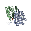
| Unit cell |
|
-Components
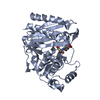
-Protein / Sugars , 2 types, 4 molecules AB
| #1: Protein | / CD Man-6-P receptor / CD-MPR / 46 kDa mannose 6-phosphate receptor / MPR 46 Mass: 17444.611 Da / Num. of mol.: 2 / Fragment: UNP residues 29-182 / Mutation: E19Q, K137M,N31Q, N57Q, N68Q, N87Q Source method: isolated from a genetically manipulated source Source: (gene. exp.) Bos taurus (cattle) / Gene: M6PR / Plasmid: pPICZalphaA / Production host:  Pichia Pastoris (fungus) / Strain (production host): x-33 / References: UniProt: P11456 Pichia Pastoris (fungus) / Strain (production host): x-33 / References: UniProt: P11456#2: Sugar | N-Acetylglucosamine Type: D-saccharide, beta linking / Mass: 221.208 Da / Num. of mol.: 2 Type: D-saccharide, beta linking / Mass: 221.208 Da / Num. of mol.: 2Source method: isolated from a genetically manipulated source Formula: C8H15NO6 |
|---|
-Non-polymers , 4 types, 200 molecules 


| #3: Chemical | Sulfate Mass: 96.063 Da / Num. of mol.: 3 / Source method: obtained synthetically / Formula: SO4 Mass: 96.063 Da / Num. of mol.: 3 / Source method: obtained synthetically / Formula: SO4#4: Chemical | ChemComp-ACT / | Acetate Mass: 59.044 Da / Num. of mol.: 1 / Source method: obtained synthetically / Formula: C2H3O2 Mass: 59.044 Da / Num. of mol.: 1 / Source method: obtained synthetically / Formula: C2H3O2#5: Chemical | ChemComp-IMD / | Imidazole Mass: 69.085 Da / Num. of mol.: 1 / Source method: obtained synthetically / Formula: C3H5N2 Mass: 69.085 Da / Num. of mol.: 1 / Source method: obtained synthetically / Formula: C3H5N2#6: Water | ChemComp-HOH / | WaterMass: 18.015 Da / Num. of mol.: 195 / Source method: isolated from a natural source / Formula: H2O |
|---|
-Experimental details
-Experiment
| Experiment | Method: X-RAY DIFFRACTION / Number of used crystals: 1 |
|---|
- Sample preparation
| Crystal | Density Matthews: 3.79 Å3/Da / Density % sol: 67.57 % |
|---|---|
| Crystal grow | Temperature: 292 K / Method: vapor diffusion, hanging drop / pH: 6.5 Details: 0.1M sodium cacodylate, 3M ammonium sulfate, pH 6.5, VAPOR DIFFUSION, HANGING DROP, temperature 292K |
-Data collection
| Diffraction | Mean temperature: 98 K | |||||||||||||||||||||||||||||||||||||||||||||||||||||||||||||||||||||||||||||
|---|---|---|---|---|---|---|---|---|---|---|---|---|---|---|---|---|---|---|---|---|---|---|---|---|---|---|---|---|---|---|---|---|---|---|---|---|---|---|---|---|---|---|---|---|---|---|---|---|---|---|---|---|---|---|---|---|---|---|---|---|---|---|---|---|---|---|---|---|---|---|---|---|---|---|---|---|---|---|
| Diffraction source | Source: ROTATING ANODE / Type: RIGAKU MICROMAX-007 / Wavelength: 1.54 Å | |||||||||||||||||||||||||||||||||||||||||||||||||||||||||||||||||||||||||||||
| Detector | Type: RIGAKU RAXIS IV++ / Detector: IMAGE PLATE / Date: Apr 15, 2009 | |||||||||||||||||||||||||||||||||||||||||||||||||||||||||||||||||||||||||||||
| Radiation | Protocol: SINGLE WAVELENGTH / Monochromatic (M) / Laue (L): M / Scattering type: x-ray | |||||||||||||||||||||||||||||||||||||||||||||||||||||||||||||||||||||||||||||
| Radiation wavelength | Wavelength: 1.54 Å / Relative weight: 1 | |||||||||||||||||||||||||||||||||||||||||||||||||||||||||||||||||||||||||||||
| Reflection twin | Operator: h,-k,-l / Fraction: 0.284 | |||||||||||||||||||||||||||||||||||||||||||||||||||||||||||||||||||||||||||||
| Reflection | Resolution: 2→50 Å / Num. obs: 35223 / % possible obs: 99.7 % / Redundancy: 5.7 % / Rmerge(I) obs: 0.068 / Χ2: 1.262 / Net I/σ(I): 15.2 | |||||||||||||||||||||||||||||||||||||||||||||||||||||||||||||||||||||||||||||
| Reflection shell |
|
- Processing
| Software |
| ||||||||||||||||||||||||||||
|---|---|---|---|---|---|---|---|---|---|---|---|---|---|---|---|---|---|---|---|---|---|---|---|---|---|---|---|---|---|
| Refinement | Method to determine structure: difference fourier Starting model: PDB entry 3K42 Resolution: 2→27.46 Å / Occupancy max: 1 / Occupancy min: 1 / σ(F): 271
| ||||||||||||||||||||||||||||
| Solvent computation | Bsol: 57.299 Å2 | ||||||||||||||||||||||||||||
| Displacement parameters | Biso max: 74.73 Å2 / Biso mean: 30.533 Å2 / Biso min: 12.51 Å2
| ||||||||||||||||||||||||||||
| Refinement step | Cycle: LAST / Resolution: 2→27.46 Å
| ||||||||||||||||||||||||||||
| Refine LS restraints |
| ||||||||||||||||||||||||||||
| Xplor file |
|
