 Movie
Movie Controller


+ Open data

- Basic information
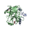
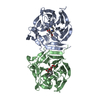
| Entry | Database: PDB / ID: 3jz1 | ||||||
|---|---|---|---|---|---|---|---|
| Title | Crystal structure of human thrombin mutant N143P in E:Na+ form | ||||||
 Components Components |
| ||||||
 Keywords Keywords |  HYDROLASE / Serine protease / Acute phase / Blood coagulation / Cleavage on pair of basic residues / Disease mutation / Disulfide bond / Gamma-carboxyglutamic acid / Glycoprotein / Kringle / Protease / Secreted / Zymogen HYDROLASE / Serine protease / Acute phase / Blood coagulation / Cleavage on pair of basic residues / Disease mutation / Disulfide bond / Gamma-carboxyglutamic acid / Glycoprotein / Kringle / Protease / Secreted / Zymogen | ||||||
| Function / homology |  Function and homology information Function and homology informationpositive regulation of lipid kinase activity / positive regulation of phospholipase C-activating G protein-coupled receptor signaling pathway / cytolysis by host of symbiont cells / thrombospondin receptor activity / Defective factor XII causes hereditary angioedema / thrombin / regulation of blood coagulation / ligand-gated ion channel signaling pathway / neutrophil-mediated killing of gram-negative bacterium / Defective F8 cleavage by thrombin ...positive regulation of lipid kinase activity / positive regulation of phospholipase C-activating G protein-coupled receptor signaling pathway / cytolysis by host of symbiont cells / thrombospondin receptor activity / Defective factor XII causes hereditary angioedema / thrombin / regulation of blood coagulation / ligand-gated ion channel signaling pathway / neutrophil-mediated killing of gram-negative bacterium / Defective F8 cleavage by thrombin / Platelet Aggregation (Plug Formation) / negative regulation of astrocyte differentiation / negative regulation of platelet activation / positive regulation of collagen biosynthetic process / negative regulation of cytokine production involved in inflammatory response / positive regulation of blood coagulation / negative regulation of fibrinolysis / Gamma-carboxylation of protein precursors / Transport of gamma-carboxylated protein precursors from the endoplasmic reticulum to the Golgi apparatus / Common Pathway of Fibrin Clot Formation / Removal of aminoterminal propeptides from gamma-carboxylated proteins / fibrinolysis / regulation of cytosolic calcium ion concentration / Intrinsic Pathway of Fibrin Clot Formation / Peptide ligand-binding receptors / positive regulation of release of sequestered calcium ion into cytosol / acute-phase response / Regulation of Complement cascade / negative regulation of proteolysis / Cell surface interactions at the vascular wall / lipopolysaccharide binding / positive regulation of receptor signaling pathway via JAK-STAT / growth factor activity / positive regulation of insulin secretion / platelet activation / response to wounding / Golgi lumen / positive regulation of protein localization to nucleus / Regulation of Insulin-like Growth Factor (IGF) transport and uptake by Insulin-like Growth Factor Binding Proteins (IGFBPs) / positive regulation of reactive oxygen species metabolic process / blood coagulation / antimicrobial humoral immune response mediated by antimicrobial peptide / Thrombin signalling through proteinase activated receptors (PARs) / heparin binding / regulation of cell shape / positive regulation of cell growth / G alpha (q) signalling events / collagen-containing extracellular matrix / blood microparticle / positive regulation of phosphatidylinositol 3-kinase/protein kinase B signal transduction / cell surface receptor signaling pathway / positive regulation of protein phosphorylation / G protein-coupled receptor signaling pathway / endoplasmic reticulum lumen / signaling receptor binding / serine-type endopeptidase activity / calcium ion binding / positive regulation of cell population proliferation / proteolysis / extracellular space / extracellular exosome / extracellular region / plasma membraneSimilarity search - Function | ||||||
| Biological species |  Homo sapiens (human) Homo sapiens (human) | ||||||
| Method | X-RAY DIFFRACTION / SYNCHROTRON / MOLECULAR REPLACEMENT / Resolution: 1.6 Å | ||||||
 Authors Authors | Niu, W. / Chen, Z. / Bush-Pelc, L.A. / Bah, A. / Gandhi, P.S. / Di Cera, E. | ||||||
 Citation Citation | Journal: J.Biol.Chem. / Year: 2009 Title: Mutant N143P reveals how Na+ activates thrombin Authors: Niu, W. / Chen, Z. / Bush-Pelc, L.A. / Bah, A. / Gandhi, P.S. / Di Cera, E. #1: Journal: J.Biol.Chem. / Year: 2004Title: Molecular dissection of Na+ binding to thrombin Authors: Pineda, A.O. / Carrell, C.J. / Bush, L.A. / Prasad, S. / Caccia, S. / Chen, Z. / Mathews, F.S. / Di Cera, E. #2: Journal: Proc.Natl.Acad.Sci.USA / Year: 2009Title: Structural identification of the pathway of long-range communication in an allosteric enzyme Authors: Gandhi, P.S. / Chen, Z. / Mathews, F.S. / Di Cera, E. | ||||||
| History |
|
- Structure visualization
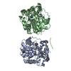
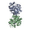
| Structure viewer | Molecule: MolmilJmol/JSmol |
|---|
- Downloads & links
-Download
| PDBx/mmCIF format | 3jz1.cif.gz | 79.2 KB | Display | PDBx/mmCIF format |
|---|---|---|---|---|
| PDB format | pdb3jz1.ent.gz | 56.6 KB | Display | PDB format |
| PDBx/mmJSON format | 3jz1.json.gz | Tree view | PDBx/mmJSON format | |
| Others |  Other downloads Other downloads |
-Validation report
| Arichive directory | https://data.pdbj.org/pub/pdb/validation_reports/jz/3jz1ftp://data.pdbj.org/pub/pdb/validation_reports/jz/3jz1 | HTTPS FTP |
|---|
-Related structure data
| Related structure data | 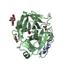 3jz2C 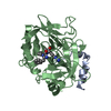 1shhS S: Starting model for refinement C: citing same article ( |
|---|---|
| Similar structure data |


-Links
 PDBj
PDBj


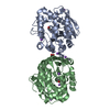
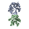
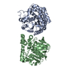
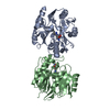
- Assembly
| Deposited unit | 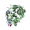
| |||||||||
|---|---|---|---|---|---|---|---|---|---|---|
| 1 | 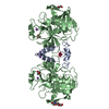
| |||||||||
| 2 |
| |||||||||
| Unit cell |
| |||||||||
| Components on special symmetry positions |
|
-Components
-Protein/peptide / Protein / Sugars , 3 types, 3 molecules AB
| #1: Protein/peptide | Mass: 4096.534 Da / Num. of mol.: 1 / Mutation: N143P Source method: isolated from a genetically manipulated source Source: (gene. exp.) Homo sapiens (human) / Gene: F2 / Production host:   Cricetulus griseus (Chinese hamster) / References: UniProt: P00734, thrombin Cricetulus griseus (Chinese hamster) / References: UniProt: P00734, thrombin |
|---|---|
| #2: Protein | Mass: 29763.234 Da / Num. of mol.: 1 Source method: isolated from a genetically manipulated source Source: (gene. exp.) Homo sapiens (human) / Gene: F2 / Production host: Cricetulus griseus (Chinese hamster) / References: UniProt: P00734, thrombin |
| #5: Sugar | ChemComp-NAG / N-Acetylglucosamine Type: D-saccharide, beta linking / Mass: 221.208 Da / Num. of mol.: 1 Type: D-saccharide, beta linking / Mass: 221.208 Da / Num. of mol.: 1Source method: isolated from a genetically manipulated source Formula: C8H15NO6 |
-Non-polymers , 4 types, 219 molecules 


| #3: Chemical |  Mass: 22.990 Da / Num. of mol.: 2 / Source method: obtained synthetically / Formula: Na Mass: 22.990 Da / Num. of mol.: 2 / Source method: obtained synthetically / Formula: Na#4: Chemical | Nitrate Mass: 62.005 Da / Num. of mol.: 2 / Source method: obtained synthetically / Formula: NO3 Mass: 62.005 Da / Num. of mol.: 2 / Source method: obtained synthetically / Formula: NO3#6: Chemical | ChemComp-GOL / | Glycerol Mass: 92.094 Da / Num. of mol.: 1 / Source method: obtained synthetically / Formula: C3H8O3 Mass: 92.094 Da / Num. of mol.: 1 / Source method: obtained synthetically / Formula: C3H8O3#7: Water | ChemComp-HOH / | WaterMass: 18.015 Da / Num. of mol.: 214 / Source method: isolated from a natural source / Formula: H2O |
|---|
-Experimental details
-Experiment
| Experiment | Method: X-RAY DIFFRACTION / Number of used crystals: 1 |
|---|
- Sample preparation
| Crystal | Density Matthews: 2.14 Å3/Da / Density % sol: 42.49 % |
|---|---|
| Crystal grow | Temperature: 295 K / Method: vapor diffusion, hanging drop / pH: 6.8 Details: 0.2M NaNO3 and 20% PEG 3350, pH 6.8, VAPOR DIFFUSION, HANGING DROP, temperature 295K |
-Data collection
| Diffraction | Mean temperature: 100 K |
|---|---|
| Diffraction source | Source: SYNCHROTRON / Site: APS  / Beamline: 14-BM-C / Wavelength: 0.9 Å / Beamline: 14-BM-C / Wavelength: 0.9 Å |
| Detector | Type: ADSC QUANTUM 315 / Detector: CCD / Date: Dec 21, 2008 |
| Radiation | Monochromator: Bent Ge(111) monochromator / Protocol: SINGLE WAVELENGTH / Monochromatic (M) / Laue (L): M / Scattering type: x-ray |
| Radiation wavelength | Wavelength: 0.9 Å / Relative weight: 1 |
| Reflection | Resolution: 1.6→40 Å / Num. all: 37456 / Num. obs: 36932 / % possible obs: 98.6 % / Observed criterion σ(F): -1 / Observed criterion σ(I): -1 / Redundancy: 4.1 % / Biso Wilson estimate: 18.5 Å2 / Rmerge(I) obs: 0.061 / Net I/σ(I): 16.9 |
| Reflection shell | Resolution: 1.6→1.66 Å / Redundancy: 3.3 % / Rmerge(I) obs: 0.32 / Mean I/σ(I) obs: 3.1 / Num. unique all: 3615 / % possible all: 97 |
- Processing
| Software |
| |||||||||||||||||||||||||
|---|---|---|---|---|---|---|---|---|---|---|---|---|---|---|---|---|---|---|---|---|---|---|---|---|---|---|
| Refinement | Method to determine structure: MOLECULAR REPLACEMENT Starting model: PDB entry 1SHH Resolution: 1.6→26.57 Å / Rfactor Rfree error: 0.005 / Data cutoff high absF: 343924.06 / Data cutoff low absF: 0 / Isotropic thermal model: RESTRAINED / Cross valid method: THROUGHOUT / σ(F): 0 / σ(I): 0 / Stereochemistry target values: Engh & Huber
| |||||||||||||||||||||||||
| Displacement parameters | Biso mean: 29.4 Å2 | |||||||||||||||||||||||||
| Refine analyze |
| |||||||||||||||||||||||||
| Refinement step | Cycle: LAST / Resolution: 1.6→26.57 Å
| |||||||||||||||||||||||||
| Refine LS restraints |
| |||||||||||||||||||||||||
| LS refinement shell | Resolution: 1.6→1.7 Å / Total num. of bins used: 6
|
