[English] 日本語
Yorodumi
- PDB-3jtc: Importance of Mg2+ in the Ca2+-Dependent Folding of the gamma-Car... -

+
Open data


ID or keywords:

Loading...

-
Basic information

Entry
Database: PDB / ID: 3jtc
TitleImportance of Mg2+ in the Ca2+-Dependent Folding of the gamma-Carboxyglutamic Acid Domains of Vitamin K-Dependent clotting and anticlotting Proteins
Components
  • Endothelial protein C receptor
  • Vitamin K-dependent protein C
KeywordsBLOOD CLOTTING / GLA (gamma-carboxyglutamic acid) residues / phospholipid binding groove / Ca ion binding / Blood coagulation / Disulfide bond / Glycoprotein / Membrane / Receptor / Transmembrane / Cleavage on pair of basic residues / Disease mutation / EGF-like domain / Gamma-carboxyglutamic acid / Hydrolase / Hydroxylation / Protease / Serine protease / Thrombophilia / Zymogen
Function / homology
Function and homology information


protein C (activated) / positive regulation of establishment of endothelial barrier / negative regulation of coagulation / negative regulation of blood coagulation / Gamma-carboxylation of protein precursors / Transport of gamma-carboxylated protein precursors from the endoplasmic reticulum to the Golgi apparatus / Common Pathway of Fibrin Clot Formation / Removal of aminoterminal propeptides from gamma-carboxylated proteins / Intrinsic Pathway of Fibrin Clot Formation / Cell surface interactions at the vascular wall ...protein C (activated) / positive regulation of establishment of endothelial barrier / negative regulation of coagulation / negative regulation of blood coagulation / Gamma-carboxylation of protein precursors / Transport of gamma-carboxylated protein precursors from the endoplasmic reticulum to the Golgi apparatus / Common Pathway of Fibrin Clot Formation / Removal of aminoterminal propeptides from gamma-carboxylated proteins / Intrinsic Pathway of Fibrin Clot Formation / Cell surface interactions at the vascular wall / Post-translational protein phosphorylation / negative regulation of inflammatory response / Golgi lumen / Regulation of Insulin-like Growth Factor (IGF) transport and uptake by Insulin-like Growth Factor Binding Proteins (IGFBPs) / blood coagulation / signaling receptor activity / endoplasmic reticulum lumen / serine-type endopeptidase activity / focal adhesion / centrosome / calcium ion binding / negative regulation of apoptotic process / perinuclear region of cytoplasm / Golgi apparatus / cell surface / endoplasmic reticulum / proteolysis / extracellular space / extracellular exosome / extracellular region / plasma membrane
Similarity search - Function
Endothelial protein C receptor / MHC-I family domain / Peptidase S1A, coagulation factor VII/IX/X/C/Z / Coagulation factor-like, Gla domain superfamily / Coagulation Factor Xa inhibitory site / EGF-type aspartate/asparagine hydroxylation site / EGF-like calcium-binding, conserved site / Calcium-binding EGF-like domain signature. / Aspartic acid and asparagine hydroxylation site. / EGF-like calcium-binding domain ...Endothelial protein C receptor / MHC-I family domain / Peptidase S1A, coagulation factor VII/IX/X/C/Z / Coagulation factor-like, Gla domain superfamily / Coagulation Factor Xa inhibitory site / EGF-type aspartate/asparagine hydroxylation site / EGF-like calcium-binding, conserved site / Calcium-binding EGF-like domain signature. / Aspartic acid and asparagine hydroxylation site. / EGF-like calcium-binding domain / Calcium-binding EGF-like domain / Vitamin K-dependent carboxylation/gamma-carboxyglutamic (GLA) domain / Gamma-carboxyglutamic acid-rich (GLA) domain / Gamma-carboxyglutamic acid-rich (GLA) domain superfamily / Vitamin K-dependent carboxylation domain. / Gla domain profile. / Domain containing Gla (gamma-carboxyglutamate) residues. / Epidermal growth factor-like domain. / EGF-like domain profile. / MHC class I-like antigen recognition-like / Murine Class I Major Histocompatibility Complex, H2-DB; Chain A, domain 1 / EGF-like domain signature 2. / EGF-like domain signature 1. / EGF-like domain / MHC class I-like antigen recognition-like / MHC class I-like antigen recognition-like superfamily / MHC classes I/II-like antigen recognition protein / Serine proteases, trypsin family, histidine active site / Serine proteases, trypsin family, serine active site / Peptidase S1A, chymotrypsin family / Serine proteases, trypsin family, histidine active site. / Serine proteases, trypsin domain profile. / Serine proteases, trypsin family, serine active site. / Trypsin-like serine protease / Serine proteases, trypsin domain / Trypsin / Peptidase S1, PA clan, chymotrypsin-like fold / Peptidase S1, PA clan / 2-Layer Sandwich / Alpha Beta
Similarity search - Domain/homology
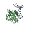
PHOSPHATIDYLETHANOLAMINE / Vitamin K-dependent protein C / Endothelial protein C receptor
Similarity search - Component
Biological speciesHOMO SAPIENS (human)
MethodX-RAY DIFFRACTION / MOLECULAR REPLACEMENT / Resolution: 1.6 Å
AuthorsBajaj, S.P. / Vadivel, K. / Agah, S. / Cascio, D. / Krishnaswamy, S. / Esmon, C. / Padmanabhan, K.
CitationJournal: J.Mol.Biol. / Year: 2013
Title: Structural and Functional Studies of gamma-Carboxyglutamic Acid Domains of Factor VIIa and Activated Protein C: Role of Magnesium at Physiological Calcium.
Authors: Vadivel, K. / Agah, S. / Messer, A.S. / Cascio, D. / Bajaj, M.S. / Krishnaswamy, S. / Esmon, C.T. / Padmanabhan, K. / Bajaj, S.P.
History
DepositionSep 11, 2009Deposition site: RCSB / Processing site: RCSB
Revision 1.0Apr 6, 2011Provider: repository / Type: Initial release
Revision 1.1Jul 13, 2011Group: Version format compliance
Revision 1.2Mar 20, 2013Group: Database references
Revision 1.3Jul 3, 2013Group: Database references
Revision 2.0Jul 29, 2020Group: Advisory / Atomic model ...Advisory / Atomic model / Data collection / Derived calculations / Non-polymer description / Structure summary
Category: atom_site / chem_comp ...atom_site / chem_comp / database_PDB_caveat / entity / pdbx_chem_comp_identifier / pdbx_entity_nonpoly / pdbx_nonpoly_scheme / pdbx_struct_conn_angle / pdbx_validate_chiral / struct_asym / struct_conn / struct_site / struct_site_gen
Item: _atom_site.B_iso_or_equiv / _atom_site.Cartn_x ..._atom_site.B_iso_or_equiv / _atom_site.Cartn_x / _atom_site.Cartn_y / _atom_site.Cartn_z / _atom_site.auth_atom_id / _atom_site.auth_comp_id / _atom_site.label_atom_id / _atom_site.label_comp_id / _atom_site.label_entity_id / _atom_site.type_symbol / _chem_comp.formula / _chem_comp.formula_weight / _chem_comp.id / _chem_comp.mon_nstd_flag / _chem_comp.name / _chem_comp.type / _pdbx_nonpoly_scheme.entity_id / _pdbx_nonpoly_scheme.mon_id / _pdbx_nonpoly_scheme.pdb_mon_id / _pdbx_struct_conn_angle.ptnr1_auth_asym_id / _pdbx_struct_conn_angle.ptnr1_auth_comp_id / _pdbx_struct_conn_angle.ptnr1_auth_seq_id / _pdbx_struct_conn_angle.ptnr1_label_asym_id / _pdbx_struct_conn_angle.ptnr1_label_atom_id / _pdbx_struct_conn_angle.ptnr1_label_comp_id / _pdbx_struct_conn_angle.ptnr1_label_seq_id / _pdbx_struct_conn_angle.ptnr2_auth_asym_id / _pdbx_struct_conn_angle.ptnr2_auth_comp_id / _pdbx_struct_conn_angle.ptnr2_auth_seq_id / _pdbx_struct_conn_angle.ptnr2_label_asym_id / _pdbx_struct_conn_angle.ptnr2_label_atom_id / _pdbx_struct_conn_angle.ptnr2_label_comp_id / _pdbx_struct_conn_angle.ptnr3_auth_asym_id / _pdbx_struct_conn_angle.ptnr3_auth_comp_id / _pdbx_struct_conn_angle.ptnr3_auth_seq_id / _pdbx_struct_conn_angle.ptnr3_label_asym_id / _pdbx_struct_conn_angle.ptnr3_label_atom_id / _pdbx_struct_conn_angle.ptnr3_label_comp_id / _pdbx_struct_conn_angle.ptnr3_label_seq_id / _pdbx_struct_conn_angle.value / _struct_asym.entity_id / _struct_conn.conn_type_id / _struct_conn.id / _struct_conn.pdbx_dist_value / _struct_conn.pdbx_leaving_atom_flag / _struct_conn.pdbx_role / _struct_conn.ptnr1_auth_asym_id / _struct_conn.ptnr1_auth_comp_id / _struct_conn.ptnr1_auth_seq_id / _struct_conn.ptnr1_label_asym_id / _struct_conn.ptnr1_label_atom_id / _struct_conn.ptnr1_label_comp_id / _struct_conn.ptnr1_label_seq_id / _struct_conn.ptnr2_auth_asym_id / _struct_conn.ptnr2_auth_comp_id / _struct_conn.ptnr2_auth_seq_id / _struct_conn.ptnr2_label_asym_id / _struct_conn.ptnr2_label_atom_id / _struct_conn.ptnr2_label_comp_id / _struct_conn.ptnr2_label_seq_id
Description: Carbohydrate remediation / Provider: repository / Type: Remediation

-
Structure visualization

Structure viewerMolecule:
MolmilJmol/JSmol

Downloads & links

-
Assembly

Deposited unit
A: Endothelial protein C receptor
B: Endothelial protein C receptor
C: Vitamin K-dependent protein C
D: Vitamin K-dependent protein C
hetero molecules


Theoretical massNumber of molelcules
Total (without water)56,10526
Polymers52,8124
Non-polymers3,29322
Water7,332407
1
A: Endothelial protein C receptor
C: Vitamin K-dependent protein C
hetero molecules


Theoretical massNumber of molelcules
Total (without water)28,05313
Polymers26,4062
Non-polymers1,64711
Water362
TypeNameSymmetry operationNumber
identity operation1_555x,y,z1
Buried area3930 Å2
ΔGint-86 kcal/mol
Surface area11410 Å2
MethodPISA
2
B: Endothelial protein C receptor
D: Vitamin K-dependent protein C
hetero molecules


Theoretical massNumber of molelcules
Total (without water)28,05313
Polymers26,4062
Non-polymers1,64711
Water362
TypeNameSymmetry operationNumber
identity operation1_555x,y,z1
Buried area4460 Å2
ΔGint-79 kcal/mol
Surface area11540 Å2
MethodPISA
Unit cell
Length a, b, c (Å)59.220, 62.360, 71.030
Angle α, β, γ (deg.)90.00, 101.81, 90.00
Int Tables number4
Space group name H-MP1211

-
Components

-
Protein / Protein/peptide / Sugars , 3 types, 10 molecules ABCD

#1: Protein Endothelial protein C receptor / / Endothelial cell protein C receptor / Activated protein C receptor / APC receptor


Mass: 22046.562 Da / Num. of mol.: 2 / Fragment: extracellular domain (UNP residues 18-210) / Source method: isolated from a natural source / Source: (natural) HOMO SAPIENS (human) / References: UniProt: Q9UNN8
#2: Protein/peptide Vitamin K-dependent protein C / Autoprothrombin IIA / Anticoagulant protein C / Blood coagulation factor XIV / Vitamin K-dependent ...Autoprothrombin IIA / Anticoagulant protein C / Blood coagulation factor XIV / Vitamin K-dependent protein C light chain / Vitamin K-dependent protein C heavy chain / Activation peptide


Mass: 4359.409 Da / Num. of mol.: 2 / Fragment: GLA domain (UNP residues 43-75) / Source method: isolated from a natural source
Details: CLEAVAGE HAPPENED DURING CRYSTALLIZATION AND THE CRYSTAL CONTAINS ONLY THE N-TERMINAL DOMAIN (GLA DOMAIN) OF PROTEIN C.
Source: (natural) HOMO SAPIENS (human) / References: UniProt: P04070, protein C (activated)
#3: Sugar
ChemComp-NAG / 2-acetamido-2-deoxy-beta-D-glucopyranose / N-Acetylglucosamine


Type: D-saccharide, beta linking / Mass: 221.208 Da / Num. of mol.: 6
Source method: isolated from a genetically manipulated source
Formula: C8H15NO6
IdentifierTypeProgram
DGlcpNAcbCONDENSED IUPAC CARBOHYDRATE SYMBOLGMML 1.0
N-acetyl-b-D-glucopyranosamineCOMMON NAMEGMML 1.0
b-D-GlcpNAcIUPAC CARBOHYDRATE SYMBOLPDB-CARE 1.0
GlcNAcSNFG CARBOHYDRATE SYMBOLGMML 1.0

-
Non-polymers , 4 types, 423 molecules

#4: Chemical ChemComp-PTY / PHOSPHATIDYLETHANOLAMINE / Phosphatidylethanolamine


Mass: 734.039 Da / Num. of mol.: 2 / Source method: obtained synthetically / Formula: C40H80NO8P / Comment: phospholipid*YM
#5: Chemical
ChemComp-MG / MAGNESIUM ION


Mass: 24.305 Da / Num. of mol.: 4 / Source method: obtained synthetically / Formula: Mg
#6: Chemical
ChemComp-CA / CALCIUM ION


Mass: 40.078 Da / Num. of mol.: 10 / Source method: obtained synthetically / Formula: Ca
#7: Water ChemComp-HOH / water / Water


Mass: 18.015 Da / Num. of mol.: 407 / Source method: isolated from a natural source / Formula: H2O

-
Details

Compound detailsCLEAVAGE HAPPENED DURING CRYSTALLIZATION AND THE CRYSTAL CONTAINS ONLY THE N-TERMINAL DOMAIN (GLA ...CLEAVAGE HAPPENED DURING CRYSTALLIZATION AND THE CRYSTAL CONTAINS ONLY THE N-TERMINAL DOMAIN (GLA DOMAIN) OF PROTEIN C.

-
Experimental details

-
Experiment

ExperimentMethod: X-RAY DIFFRACTION / Number of used crystals: 1

-
Sample preparation

CrystalDensity Matthews: 2.43 Å3/Da / Density % sol: 49.39 %
Crystal growMethod: vapor diffusion / pH: 7
Details: PEG 400, potassium chloride, magnesium chloride, calcium chloride, HEPES, PH 7.0, VAPOR DIFFUSION

-
Data collection

DiffractionMean temperature: 110 K
Diffraction sourceSource: ROTATING ANODE / Type: RIGAKU RU300
DetectorType: MARRESEARCH / Detector: IMAGE PLATE / Date: Apr 25, 2001 / Details: MIRRORS
RadiationProtocol: SINGLE WAVELENGTH / Monochromatic (M) / Laue (L): M / Scattering type: x-ray
Radiation wavelengthRelative weight: 1
ReflectionResolution: 1.55→30 Å / Num. obs: 66375 / % possible obs: 90.1 % / Observed criterion σ(I): -3 / Redundancy: 4 % / Rsym value: 0.041 / Net I/σ(I): 27
Reflection shellResolution: 1.55→1.61 Å / Redundancy: 4 % / Rmerge(I) obs: 0.455 / Mean I/σ(I) obs: 27 / Rsym value: 0.043 / % possible all: 91

-
Processing

Software
NameVersionClassificationNB
REFMAC5.2.0019refinement
PDB_EXTRACT3.005data extraction
HKL-2000data reduction
SCALEPACKdata scaling
RefinementMethod to determine structure: MOLECULAR REPLACEMENT / Resolution: 1.6→23.22 Å / Cor.coef. Fo:Fc: 0.962 / Cor.coef. Fo:Fc free: 0.948 / Occupancy max: 1 / Occupancy min: 0.5 / Cross valid method: THROUGHOUT / ESU R: 0.102 / ESU R Free: 0.102 / Stereochemistry target values: MAXIMUM LIKELIHOOD / Details: HYDROGENS HAVE BEEN ADDED IN THE RIDING POSITIONS
RfactorNum. reflection% reflectionSelection details
Rfree0.22648 3024 5.1 %RANDOM
Rwork0.19066 ---
obs0.19246 56575 89.13 %-
Solvent computationIon probe radii: 0.8 Å / Shrinkage radii: 0.8 Å / VDW probe radii: 1.4 Å / Solvent model: MASK
Displacement parametersBiso mean: 25.75 Å2
Baniso -1Baniso -2Baniso -3
1-0.13 Å20 Å21.12 Å2
2--0.87 Å20 Å2
3----0.55 Å2
Refinement stepCycle: LAST / Resolution: 1.6→23.22 Å
ProteinNucleic acidLigandSolventTotal
Num. atoms3422 0 198 407 4027
Refine LS restraints
Refine-IDTypeDev idealDev ideal targetNumber
X-RAY DIFFRACTIONr_bond_refined_d1.450.0213695
X-RAY DIFFRACTIONr_bond_other_d
X-RAY DIFFRACTIONr_angle_refined_deg2.5662.0335012
X-RAY DIFFRACTIONr_angle_other_deg
X-RAY DIFFRACTIONr_dihedral_angle_1_deg6.2495407
X-RAY DIFFRACTIONr_dihedral_angle_2_deg35.98223.064173
X-RAY DIFFRACTIONr_dihedral_angle_3_deg15.45715530
X-RAY DIFFRACTIONr_dihedral_angle_4_deg22.4721528
X-RAY DIFFRACTIONr_chiral_restr0.1730.2516
X-RAY DIFFRACTIONr_gen_planes_refined0.0320.0252841
X-RAY DIFFRACTIONr_gen_planes_other
X-RAY DIFFRACTIONr_nbd_refined0.2450.21724
X-RAY DIFFRACTIONr_nbd_other
X-RAY DIFFRACTIONr_nbtor_refined0.3040.22505
X-RAY DIFFRACTIONr_nbtor_other
X-RAY DIFFRACTIONr_xyhbond_nbd_refined0.1260.2322
X-RAY DIFFRACTIONr_xyhbond_nbd_other
X-RAY DIFFRACTIONr_metal_ion_refined0.1550.235
X-RAY DIFFRACTIONr_metal_ion_other
X-RAY DIFFRACTIONr_symmetry_vdw_refined0.210.285
X-RAY DIFFRACTIONr_symmetry_vdw_other
X-RAY DIFFRACTIONr_symmetry_hbond_refined0.1180.224
X-RAY DIFFRACTIONr_symmetry_hbond_other
X-RAY DIFFRACTIONr_symmetry_metal_ion_refined
X-RAY DIFFRACTIONr_symmetry_metal_ion_other
X-RAY DIFFRACTIONr_mcbond_it1.611.52050
X-RAY DIFFRACTIONr_mcbond_other
X-RAY DIFFRACTIONr_mcangle_it2.62323315
X-RAY DIFFRACTIONr_scbond_it3.61231645
X-RAY DIFFRACTIONr_scangle_it5.3714.51697
X-RAY DIFFRACTIONr_rigid_bond_restr
X-RAY DIFFRACTIONr_sphericity_free
X-RAY DIFFRACTIONr_sphericity_bonded
LS refinement shellResolution: 1.6→1.641 Å / Total num. of bins used: 20
RfactorNum. reflection% reflection
Rfree0.487 139 -
Rwork0.414 2850 -
obs--61.09 %

+
About Yorodumi

-
News

-
Feb 9, 2022. New format data for meta-information of EMDB entries

New format data for meta-information of EMDB entries

  • Version 3 of the EMDB header file is now the official format.
  • The previous official version 1.9 will be removed from the archive.

Related info.:EMDB header

External links:wwPDB to switch to version 3 of the EMDB data model

-
Aug 12, 2020. Covid-19 info

Covid-19 info

URL: https://pdbj.org/emnavi/covid19.php

New page: Covid-19 featured information page in EM Navigator.

Related info.:Covid-19 info / Mar 5, 2020. Novel coronavirus structure data

+
Mar 5, 2020. Novel coronavirus structure data

Novel coronavirus structure data

Related info.:Yorodumi Speices / Aug 12, 2020. Covid-19 info

External links:COVID-19 featured content - PDBj / Molecule of the Month (242):Coronavirus Proteases

+
Jan 31, 2019. EMDB accession codes are about to change! (news from PDBe EMDB page)

EMDB accession codes are about to change! (news from PDBe EMDB page)

  • The allocation of 4 digits for EMDB accession codes will soon come to an end. Whilst these codes will remain in use, new EMDB accession codes will include an additional digit and will expand incrementally as the available range of codes is exhausted. The current 4-digit format prefixed with “EMD-” (i.e. EMD-XXXX) will advance to a 5-digit format (i.e. EMD-XXXXX), and so on. It is currently estimated that the 4-digit codes will be depleted around Spring 2019, at which point the 5-digit format will come into force.
  • The EM Navigator/Yorodumi systems omit the EMD- prefix.

Related info.:Q: What is EMD? / ID/Accession-code notation in Yorodumi/EM Navigator

External links:EMDB Accession Codes are Changing Soon! / Contact to PDBj

+
Jul 12, 2017. Major update of PDB

Major update of PDB

  • wwPDB released updated PDB data conforming to the new PDBx/mmCIF dictionary.
  • This is a major update changing the version number from 4 to 5, and with Remediation, in which all the entries are updated.
  • In this update, many items about electron microscopy experimental information are reorganized (e.g. em_software).
  • Now, EM Navigator and Yorodumi are based on the updated data.

External links:wwPDB Remediation / Enriched Model Files Conforming to OneDep Data Standards Now Available in the PDB FTP Archive

-
Yorodumi

Thousand views of thousand structures

  • Yorodumi is a browser for structure data from EMDB, PDB, SASBDB, etc.
  • This page is also the successor to EM Navigator detail page, and also detail information page/front-end page for Omokage search.
  • The word "yorodu" (or yorozu) is an old Japanese word meaning "ten thousand". "mi" (miru) is to see.

Related info.:EMDB / PDB / SASBDB / Comparison of 3 databanks / Yorodumi Search / Aug 31, 2016. New EM Navigator & Yorodumi / Yorodumi Papers / Jmol/JSmol / Function and homology information / Changes in new EM Navigator and Yorodumi

Read more