 Movie
Movie Controller

[English] 日本語
 Yorodumi
- PDB-2ia4: Crystal structure of Novel amino acid binding protein from Shigel... -
+ Open data

- Basic information
| Entry | Database: PDB / ID: 2ia4 | ||||||
|---|---|---|---|---|---|---|---|
| Title | Crystal structure of Novel amino acid binding protein from Shigella flexneri | ||||||
 Components Components | Putative periplasmic binding transport protein | ||||||
 Keywords Keywords |  PROTEIN TRANSPORT / beta-alpha mixture PROTEIN TRANSPORT / beta-alpha mixture | ||||||
| Function / homology |  Function and homology information Function and homology informationBacterial periplasmic substrate-binding proteins / Bacterial extracellular solute-binding proteins, family 3 / Solute-binding protein family 3/N-terminal domain of MltF / Periplasmic binding protein-like II / D-Maltodextrin-Binding Protein; domain 2 / 3-Layer(aba) Sandwich / Alpha Beta Similarity search - Domain/homology | ||||||
| Biological species |  Shigella flexneri 2a str. 301 (bacteria) Shigella flexneri 2a str. 301 (bacteria) | ||||||
| Method | X-RAY DIFFRACTION / SYNCHROTRON / MAD / Resolution: 1.5 Å | ||||||
 Authors Authors | Fan, C.P. / Wang, D.C. | ||||||
 Citation Citation | Journal: To be Published Title: Structural and functional study of novel amino acid binding protein from Shigella flexneri Authors: Fan, C.P. / Wang, D.C. | ||||||
| History |
|
- Structure visualization
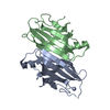
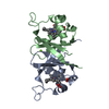
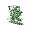
| Structure viewer | Molecule: MolmilJmol/JSmol |
|---|
- Downloads & links
-Download
| PDBx/mmCIF format | 2ia4.cif.gz | 130.8 KB | Display | PDBx/mmCIF format |
|---|---|---|---|---|
| PDB format | pdb2ia4.ent.gz | 107.7 KB | Display | PDB format |
| PDBx/mmJSON format | 2ia4.json.gz | Tree view | PDBx/mmJSON format | |
| Others |  Other downloads Other downloads |
-Validation report
| Arichive directory | https://data.pdbj.org/pub/pdb/validation_reports/ia/2ia4ftp://data.pdbj.org/pub/pdb/validation_reports/ia/2ia4 | HTTPS FTP |
|---|
-Related structure data
| Similar structure data |
|---|


-Links
 PDBj
PDBj

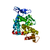
- Assembly
| Deposited unit | 
| ||||||||
|---|---|---|---|---|---|---|---|---|---|
| 1 | 
| ||||||||
| 2 | 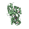
| ||||||||
| Unit cell |
|
-Components
| #1: Protein | Mass: 32531.609 Da / Num. of mol.: 2 / Fragment: residues 1-279 Source method: isolated from a genetically manipulated source Source: (gene. exp.) Shigella flexneri 2a str. 301 (bacteria)Species: Shigella flexneri / Strain: 2a 301 / Plasmid: pET22b(+) / Production host: Escherichia coli (E. coli) / Strain (production host): B834(DE3) / References: UniProt: Q83S74, UniProt: A0A0H2UXX1*PLUS#2: Chemical | Tris  Mass: 122.143 Da / Num. of mol.: 2 / Source method: obtained synthetically / Formula: C4H12NO3 / Comment: pH buffer*YM Mass: 122.143 Da / Num. of mol.: 2 / Source method: obtained synthetically / Formula: C4H12NO3 / Comment: pH buffer*YM#3: Chemical | Glutamic acid  Type: L-peptide linking / Mass: 147.129 Da / Num. of mol.: 2 / Source method: obtained synthetically / Formula: C5H9NO4 Type: L-peptide linking / Mass: 147.129 Da / Num. of mol.: 2 / Source method: obtained synthetically / Formula: C5H9NO4#4: Water | ChemComp-HOH / | Water Mass: 18.015 Da / Num. of mol.: 676 / Source method: isolated from a natural source / Formula: H2O Mass: 18.015 Da / Num. of mol.: 676 / Source method: isolated from a natural source / Formula: H2O |
|---|
-Experimental details
-Experiment
| Experiment | Method: X-RAY DIFFRACTION / Number of used crystals: 1 |
|---|
- Sample preparation
| Crystal | Density Matthews: 1.99 Å3/Da / Density % sol: 38.1 % |
|---|---|
| Crystal grow | Temperature: 293 K / Method: vapor diffusion, hanging drop / pH: 8.5 Details: trimethylamine N-oxide dihydrate, Polyethylene Glycol, Monomethyl ether 2000, pH 8.50, VAPOR DIFFUSION, HANGING DROP, temperature 293K |
-Data collection
| Diffraction | Mean temperature: 95 K | ||||||||||||
|---|---|---|---|---|---|---|---|---|---|---|---|---|---|
| Diffraction source | Source: SYNCHROTRON / Site: BSRF  / Beamline: 3W1A / Wavelength: 0.9500, 0.9794, 0.9799 / Beamline: 3W1A / Wavelength: 0.9500, 0.9794, 0.9799 | ||||||||||||
| Detector | Type: MAR CCD 165 mm / Detector: CCD / Date: Jun 19, 2005 | ||||||||||||
| Radiation | Protocol: MAD / Monochromatic (M) / Laue (L): M / Scattering type: x-ray | ||||||||||||
| Radiation wavelength |
| ||||||||||||
| Reflection | Resolution: 1.5→50 Å / Num. all: 160587 / Num. obs: 154166 / % possible obs: 99.9 % / Observed criterion σ(F): 1 / Observed criterion σ(I): 2 / Redundancy: 6.2 % / Biso Wilson estimate: 11.2 Å2 / Rmerge(I) obs: 0.066 / Rsym value: 0.066 / Net I/σ(I): 28.7 | ||||||||||||
| Reflection shell | Resolution: 1.5→1.55 Å / Redundancy: 5.6 % / Rmerge(I) obs: 0.286 / Mean I/σ(I) obs: 4 / Num. unique all: 8038 / Rsym value: 0.286 / % possible all: 99.4 |
- Processing
| Software |
| |||||||||||||||||||||||||
|---|---|---|---|---|---|---|---|---|---|---|---|---|---|---|---|---|---|---|---|---|---|---|---|---|---|---|
| Refinement | Method to determine structure: MAD / Resolution: 1.5→33.36 Å / Rfactor Rfree error: 0.002 / Data cutoff high absF: 261723.36 / Data cutoff low absF: 0 / Isotropic thermal model: RESTRAINED / Cross valid method: THROUGHOUT / σ(F): 0 / σ(I): 0 / Stereochemistry target values: Engh & Huber
| |||||||||||||||||||||||||
| Solvent computation | Solvent model: FLAT MODEL / Bsol: 44.9813 Å2 / ksol: 0.338119 e/Å3 | |||||||||||||||||||||||||
| Displacement parameters | Biso mean: 15.5 Å2
| |||||||||||||||||||||||||
| Refine analyze |
| |||||||||||||||||||||||||
| Refinement step | Cycle: LAST / Resolution: 1.5→33.36 Å
| |||||||||||||||||||||||||
| Refine LS restraints |
| |||||||||||||||||||||||||
| LS refinement shell | Resolution: 1.5→1.59 Å / Rfactor Rfree error: 0.006 / Total num. of bins used: 6
| |||||||||||||||||||||||||
| Xplor file |
|
