 Movie
Movie Controller

[English] 日本語
 Yorodumi
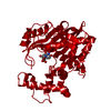
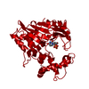
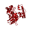
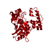
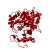
- PDB-3ijz: Lactobacillus casei Thymidylate Synthase ternary complex with dUM... -
+ Open data

- Basic information
| Entry | Database: PDB / ID: 3ijz | ||||||
|---|---|---|---|---|---|---|---|
| Title | Lactobacillus casei Thymidylate Synthase ternary complex with dUMP and Pthalimidic derivative 15C | ||||||
 Components Components | Thymidylate synthase | ||||||
 Keywords Keywords | TRANSFERASE / nucleotide synthase / Methyltransferase / Nucleotide biosynthesis | ||||||
| Function / homology |  Function and homology informationthymidylate synthase / thymidylate synthase activity / dTMP biosynthetic process / dTTP biosynthetic process / methylation / cytosol Function and homology informationthymidylate synthase / thymidylate synthase activity / dTMP biosynthetic process / dTTP biosynthetic process / methylation / cytosolSimilarity search - Function | ||||||
| Biological species |  Lactobacillus casei (bacteria) Lactobacillus casei (bacteria) | ||||||
| Method | X-RAY DIFFRACTION / SYNCHROTRON / MOLECULAR REPLACEMENT / Resolution: 2.21 Å | ||||||
 Authors Authors | Pozzi, C. / Cancian, L. / Leone, R. / Luciani, R. / Ferrari, S. / Mangani, S. / Costi, M.P. | ||||||
 Citation Citation | Journal: J.Med.Chem. / Year: 2011 Title: Identification of the binding modes of N-phenylphthalimides inhibiting bacterial thymidylate synthase through X-ray crystallography screening Authors: Mangani, S. / Cancian, L. / Leone, R. / Pozzi, C. / Lazzari, S. / Luciani, R. / Ferrari, S. / Costi, M.P. | ||||||
| History |
|
- Structure visualization
| Structure viewer | Molecule: MolmilJmol/JSmol |
|---|
- Downloads & links
-Download
| PDBx/mmCIF format | 3ijz.cif.gz | 82.7 KB | Display | PDBx/mmCIF format |
|---|---|---|---|---|
| PDB format | pdb3ijz.ent.gz | 61.4 KB | Display | PDB format |
| PDBx/mmJSON format | 3ijz.json.gz | Tree view | PDBx/mmJSON format | |
| Others |  Other downloads Other downloads |
-Validation report
| Arichive directory | https://data.pdbj.org/pub/pdb/validation_reports/ij/3ijzftp://data.pdbj.org/pub/pdb/validation_reports/ij/3ijz | HTTPS FTP |
|---|
-Related structure data
| Related structure data |  3bnzC 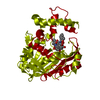 3byxC  3bz0C  3c06C 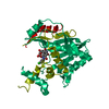 3c0aC 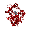 3ik0C 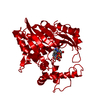 3ik1C 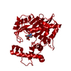 2tdmS C: citing same article ( S: Starting model for refinement |
|---|---|
| Similar structure data |

-Links
 PDBj
PDBj- Assembly
| Deposited unit | 
| ||||||||
|---|---|---|---|---|---|---|---|---|---|
| 1 | 
| ||||||||
| Unit cell |
|
-Components
| #1: Protein | / TS / TSase Mass: 36630.453 Da / Num. of mol.: 1 Source method: isolated from a genetically manipulated source Source: (gene. exp.) Lactobacillus casei (bacteria) / Production host: Escherichia coli (E. coli) / References: UniProt: P00469, thymidylate synthase |
|---|---|
| #2: Chemical | ChemComp-15C /   Mass: 279.290 Da / Num. of mol.: 1 / Source method: obtained synthetically / Formula: C17H13NO3 Mass: 279.290 Da / Num. of mol.: 1 / Source method: obtained synthetically / Formula: C17H13NO3 |
| #3: Chemical | ChemComp-UMP / Deoxyuridine monophosphate  Mass: 308.182 Da / Num. of mol.: 1 / Source method: obtained synthetically / Formula: C9H13N2O8P Mass: 308.182 Da / Num. of mol.: 1 / Source method: obtained synthetically / Formula: C9H13N2O8P |
| #4: Water | ChemComp-HOH / Water Mass: 18.015 Da / Num. of mol.: 147 / Source method: isolated from a natural source / Formula: H2O Mass: 18.015 Da / Num. of mol.: 147 / Source method: isolated from a natural source / Formula: H2O |
-Experimental details
-Experiment
| Experiment | Method: X-RAY DIFFRACTION / Number of used crystals: 1 |
|---|
- Sample preparation
| Crystal | Density Matthews: 2.67 Å3/Da / Density % sol: 53.98 % |
|---|---|
| Crystal grow | Temperature: 298 K / Method: vapor diffusion, hanging drop / pH: 6.8 Details: 10% saturated Ammonium sulfate, 1mM DTT, 20mM Potassium phosphate, pH 6.8, VAPOR DIFFUSION, HANGING DROP, temperature 298.0K |
-Data collection
| Diffraction | Mean temperature: 100 K |
|---|---|
| Diffraction source | Source: SYNCHROTRON / Site: ESRF  / Beamline: ID23-1 / Wavelength: 0.983 Å / Beamline: ID23-1 / Wavelength: 0.983 Å |
| Detector | Type: ADSC QUANTUM 315r / Detector: CCD / Date: Oct 27, 2008 |
| Radiation | Monochromator: Silicon (111) channel / Protocol: SINGLE WAVELENGTH / Monochromatic (M) / Laue (L): M / Scattering type: x-ray |
| Radiation wavelength | Wavelength: 0.983 Å / Relative weight: 1 |
| Reflection | Resolution: 2.21→74.95 Å / Num. obs: 20713 / % possible obs: 98.2 % / Observed criterion σ(F): 1 / Observed criterion σ(I): 2 / Redundancy: 16.5 % / Biso Wilson estimate: 35.031 Å2 / Rmerge(I) obs: 0.093 / Rsym value: 0.093 / Net I/σ(I): 29.6 |
| Reflection shell | Resolution: 2.21→2.33 Å / Redundancy: 16.4 % / Rmerge(I) obs: 0.356 / Mean I/σ(I) obs: 7.1 / Num. unique all: 2825 / Rsym value: 0.356 / % possible all: 95.3 |
- Processing
| Software |
| |||||||||||||||||||||||||||||||||||||||||||||||||||||||||||||||||
|---|---|---|---|---|---|---|---|---|---|---|---|---|---|---|---|---|---|---|---|---|---|---|---|---|---|---|---|---|---|---|---|---|---|---|---|---|---|---|---|---|---|---|---|---|---|---|---|---|---|---|---|---|---|---|---|---|---|---|---|---|---|---|---|---|---|---|
| Refinement | Method to determine structure: MOLECULAR REPLACEMENT Starting model: PDB ENTRY 2TDM Resolution: 2.21→67.25 Å / Cor.coef. Fo:Fc: 0.932 / Cor.coef. Fo:Fc free: 0.913 / SU B: 5.5 / SU ML: 0.143 / Cross valid method: THROUGHOUT / σ(I): 2 / ESU R: 0.266 / ESU R Free: 0.215 / Stereochemistry target values: MAXIMUM LIKELIHOOD
| |||||||||||||||||||||||||||||||||||||||||||||||||||||||||||||||||
| Solvent computation | Ion probe radii: 0.8 Å / Shrinkage radii: 0.8 Å / VDW probe radii: 1.4 Å / Solvent model: MASK | |||||||||||||||||||||||||||||||||||||||||||||||||||||||||||||||||
| Displacement parameters | Biso mean: 37.059 Å2
| |||||||||||||||||||||||||||||||||||||||||||||||||||||||||||||||||
| Refine analyze |
| |||||||||||||||||||||||||||||||||||||||||||||||||||||||||||||||||
| Refinement step | Cycle: LAST / Resolution: 2.21→67.25 Å
| |||||||||||||||||||||||||||||||||||||||||||||||||||||||||||||||||
| Refine LS restraints |
| |||||||||||||||||||||||||||||||||||||||||||||||||||||||||||||||||
| LS refinement shell | Resolution: 2.21→2.267 Å / Total num. of bins used: 20
|
