 Movie
Movie Controller


+ Open data

- Basic information
| Entry | Database: PDB / ID: 2tdm | |||||||||
|---|---|---|---|---|---|---|---|---|---|---|
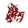
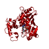
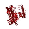
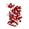
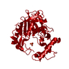
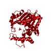
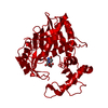
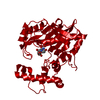
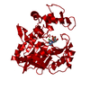
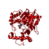
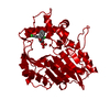
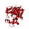
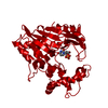
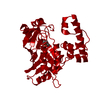
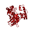
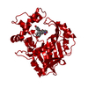
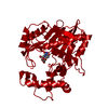
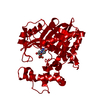
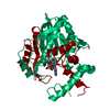
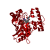
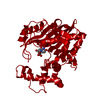
| Title | STRUCTURE OF THYMIDYLATE SYNTHASE | |||||||||
 Components Components | THYMIDYLATE SYNTHASE | |||||||||
 Keywords Keywords | METHYLTRANSFERASE / TRANSFERASE | |||||||||
| Function / homology |  Function and homology informationthymidylate synthase / thymidylate synthase activity / dTMP biosynthetic process / dTTP biosynthetic process / methylation / cytosol Function and homology informationthymidylate synthase / thymidylate synthase activity / dTMP biosynthetic process / dTTP biosynthetic process / methylation / cytosolSimilarity search - Function | |||||||||
| Biological species |  Lactobacillus casei (bacteria) Lactobacillus casei (bacteria) | |||||||||
| Method | X-RAY DIFFRACTION / Resolution: 2.55 Å | |||||||||
 Authors Authors | Finer-Moore, J. / Stroud, R.M. | |||||||||
 Citation Citation | Journal: J.Mol.Biol. / Year: 1993 Title: Refined structures of substrate-bound and phosphate-bound thymidylate synthase from Lactobacillus casei. Authors: Finer-Moore, J. / Fauman, E.B. / Foster, P.G. / Perry, K.M. / Santi, D.V. / Stroud, R.M. #1: Journal: Protein Eng. / Year: 1996Title: Contribution of a Salt Bridge to Binding Affinity and Dump Orientation to Catalytic Rate: Mutation of a Substrate-Binding Arginine in Thymidylate Synthase Authors: Finer-Moore, J.S. / Fauman, E.B. / Morse, R.J. / Santi, D.V. / Stroud, R.M. | |||||||||
| History |
|
- Structure visualization
| Structure viewer | Molecule: MolmilJmol/JSmol |
|---|
- Downloads & links
-Download
| PDBx/mmCIF format | 2tdm.cif.gz | 77.9 KB | Display | PDBx/mmCIF format |
|---|---|---|---|---|
| PDB format | pdb2tdm.ent.gz | 59 KB | Display | PDB format |
| PDBx/mmJSON format | 2tdm.json.gz | Tree view | PDBx/mmJSON format | |
| Others |  Other downloads Other downloads |
-Validation report
| Arichive directory | https://data.pdbj.org/pub/pdb/validation_reports/td/2tdmftp://data.pdbj.org/pub/pdb/validation_reports/td/2tdm | HTTPS FTP |
|---|
-Related structure data


-Links
 PDBj
PDBj- Assembly
| Deposited unit | 
| ||||||||
|---|---|---|---|---|---|---|---|---|---|
| 1 | 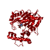
| ||||||||
| Unit cell |
|
-Components
| #1: Protein | Mass: 36630.453 Da / Num. of mol.: 1 Source method: isolated from a genetically manipulated source Source: (gene. exp.) Lactobacillus casei (bacteria)Description: STRAIN CHI-2913 LACKS A THYMIDYLATE SYNTHASE GENE Plasmid: PKPTSD / Production host: Escherichia coli (E. coli) / Strain (production host): CHI-2913 / References: UniProt: P00469, thymidylate synthase |
|---|---|
| #2: Chemical | ChemComp-UMP / Deoxyuridine monophosphate  Mass: 308.182 Da / Num. of mol.: 1 / Source method: obtained synthetically / Formula: C9H13N2O8P Mass: 308.182 Da / Num. of mol.: 1 / Source method: obtained synthetically / Formula: C9H13N2O8P |
| #3: Water | ChemComp-HOH / Water Mass: 18.015 Da / Num. of mol.: 47 / Source method: isolated from a natural source / Formula: H2O Mass: 18.015 Da / Num. of mol.: 47 / Source method: isolated from a natural source / Formula: H2O |
-Experimental details
-Experiment
| Experiment | Method: X-RAY DIFFRACTION / Number of used crystals: 1 |
|---|
- Sample preparation
| Crystal | Density Matthews: 2.92 Å3/Da / Density % sol: 55 % | ||||||||||||||||||||
|---|---|---|---|---|---|---|---|---|---|---|---|---|---|---|---|---|---|---|---|---|---|
| Crystal grow | pH: 7.4 / Details: pH 7.4 | ||||||||||||||||||||
| Crystal grow | *PLUS Method: vapor diffusion | ||||||||||||||||||||
| Components of the solutions | *PLUS
|
-Data collection
| Diffraction | Mean temperature: 295 K |
|---|---|
| Diffraction source | Source: ROTATING ANODE / Type: RIGAKU RUH2R / Wavelength: 1.5418 |
| Detector | Type: SIEMENS / Detector: AREA DETECTOR / Date: Nov 1, 1990 |
| Radiation | Monochromator: GRAPHITE(002) / Monochromatic (M) / Laue (L): M / Scattering type: x-ray |
| Radiation wavelength | Wavelength: 1.5418 Å / Relative weight: 1 |
| Reflection | Resolution: 2.55→50 Å / Num. obs: 12591 / % possible obs: 83 % / Observed criterion σ(I): 0 / Redundancy: 3.7 % / Rsym value: 0.156 / Net I/σ(I): 9.4 |
| Reflection shell | Resolution: 2.55→2.7 Å / Redundancy: 2 % / Mean I/σ(I) obs: 0.8 / Rsym value: 0.52 / % possible all: 65 |
| Reflection | *PLUS Num. measured all: 46338 / Rmerge(I) obs: 0.156 |
| Reflection shell | *PLUS % possible obs: 65 % / Rmerge(I) obs: 0.52 |
- Processing
| Software |
| ||||||||||||||||||||||||||||||||||||||||||||||||||||||||||||
|---|---|---|---|---|---|---|---|---|---|---|---|---|---|---|---|---|---|---|---|---|---|---|---|---|---|---|---|---|---|---|---|---|---|---|---|---|---|---|---|---|---|---|---|---|---|---|---|---|---|---|---|---|---|---|---|---|---|---|---|---|---|
| Refinement | Resolution: 2.55→7 Å / σ(F): 2 Details: THE SIDE CHAIN OF ARG 23 IS IN TWO HALF-OCCUPIED CONFORMATIONS IN THE CRYSTAL STRUCTURE.
| ||||||||||||||||||||||||||||||||||||||||||||||||||||||||||||
| Displacement parameters | Biso mean: 31 Å2 | ||||||||||||||||||||||||||||||||||||||||||||||||||||||||||||
| Refinement step | Cycle: LAST / Resolution: 2.55→7 Å
| ||||||||||||||||||||||||||||||||||||||||||||||||||||||||||||
| Refine LS restraints |
| ||||||||||||||||||||||||||||||||||||||||||||||||||||||||||||
| Xplor file |
| ||||||||||||||||||||||||||||||||||||||||||||||||||||||||||||
| Software | *PLUS Name: X-PLOR / Classification: refinement | ||||||||||||||||||||||||||||||||||||||||||||||||||||||||||||
| Refinement | *PLUS | ||||||||||||||||||||||||||||||||||||||||||||||||||||||||||||
| Solvent computation | *PLUS | ||||||||||||||||||||||||||||||||||||||||||||||||||||||||||||
| Displacement parameters | *PLUS | ||||||||||||||||||||||||||||||||||||||||||||||||||||||||||||
| Refine LS restraints | *PLUS
|
