 Movie
Movie Controller

[English] 日本語
 Yorodumi
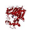
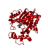
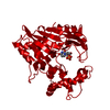
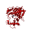
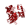
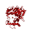
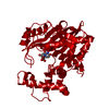
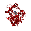
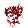
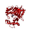
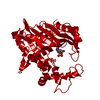
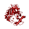
- PDB-2g86: L. casei thymidylate synthase Y261F in complex with substrate, dUMP -
+ Open data

- Basic information
| Entry | Database: PDB / ID: 2g86 | ||||||
|---|---|---|---|---|---|---|---|
| Title | L. casei thymidylate synthase Y261F in complex with substrate, dUMP | ||||||
 Components Components | Thymidylate synthase | ||||||
 Keywords Keywords | TRANSFERASE / dUMP-binding residue / dUMP complex / thymidylate synthase mutant | ||||||
| Function / homology |  Function and homology informationthymidylate synthase / thymidylate synthase activity / dTMP biosynthetic process / dTTP biosynthetic process / methylation / cytosol Function and homology informationthymidylate synthase / thymidylate synthase activity / dTMP biosynthetic process / dTTP biosynthetic process / methylation / cytosolSimilarity search - Function | ||||||
| Biological species |  Lactobacillus casei (bacteria) Lactobacillus casei (bacteria) | ||||||
| Method | X-RAY DIFFRACTION / FOURIER SYNTHESIS / Resolution: 2.4 Å | ||||||
 Authors Authors | Finer-Moore, J.S. / Stroud, R.M. | ||||||
 Citation Citation | Journal: Biochemistry / Year: 2006 Title: The role of protein dynamics in thymidylate synthase catalysis: variants of conserved 2'-deoxyuridine 5'-monophosphate (dUMP)-binding Tyr-261 Authors: Newby, Z. / Lee, T.T. / Morse, R.J. / Liu, Y. / Liu, L. / Venkatraman, P. / Santi, D.V. / Finer-Moore, J.S. / Stroud, R.M. | ||||||
| History |
|
- Structure visualization
| Structure viewer | Molecule: MolmilJmol/JSmol |
|---|
- Downloads & links
-Download
| PDBx/mmCIF format | 2g86.cif.gz | 79.2 KB | Display | PDBx/mmCIF format |
|---|---|---|---|---|
| PDB format | pdb2g86.ent.gz | 60 KB | Display | PDB format |
| PDBx/mmJSON format | 2g86.json.gz | Tree view | PDBx/mmJSON format | |
| Others |  Other downloads Other downloads |
-Validation report
| Arichive directory | https://data.pdbj.org/pub/pdb/validation_reports/g8/2g86ftp://data.pdbj.org/pub/pdb/validation_reports/g8/2g86 | HTTPS FTP |
|---|
-Related structure data

-Links
 PDBj
PDBj- Assembly
| Deposited unit | 
| ||||||||
|---|---|---|---|---|---|---|---|---|---|
| 1 | 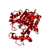
| ||||||||
| Unit cell |
| ||||||||
| Details | Biological assembly is a dimer. Second half of the dimer is generated by the crystallographic two-fold axis according to the following transformation: -x+y, y, -1/2-z |
-Components
| #1: Protein | / TS / TSase Mass: 36614.453 Da / Num. of mol.: 1 / Mutation: Y261F Source method: isolated from a genetically manipulated source Source: (gene. exp.) Lactobacillus casei (bacteria) / Gene: thyA / Plasmid: pSCTS9 / Production host: Escherichia coli (E. coli) / Strain (production host): Chi2913recA / References: UniProt: P00469, thymidylate synthase |
|---|---|
| #2: Chemical | ChemComp-UMP / Deoxyuridine monophosphate  Mass: 308.182 Da / Num. of mol.: 1 / Source method: obtained synthetically / Formula: C9H13N2O8P Mass: 308.182 Da / Num. of mol.: 1 / Source method: obtained synthetically / Formula: C9H13N2O8P |
| #3: Water | ChemComp-HOH / Water Mass: 18.015 Da / Num. of mol.: 91 / Source method: isolated from a natural source / Formula: H2O Mass: 18.015 Da / Num. of mol.: 91 / Source method: isolated from a natural source / Formula: H2O |
-Experimental details
-Experiment
| Experiment | Method: X-RAY DIFFRACTION / Number of used crystals: 1 |
|---|
- Sample preparation
| Crystal | Density Matthews: 2.94 Å3/Da / Density % sol: 58.15 % |
|---|---|
| Crystal grow | Temperature: 298 K / Method: vapor diffusion, hanging drop / pH: 7 Details: vapor diffusion against 20mM KPO4 buffer; protein solution in same buffer with 15-20mM ammonium sulfate, DTT, 38 mMdUMP, pH 7.0, VAPOR DIFFUSION, HANGING DROP, temperature 298.0K |
-Data collection
| Diffraction | Mean temperature: 298 K |
|---|---|
| Diffraction source | Source: ROTATING ANODE / Type: RIGAKU / Wavelength: 1.54178 Å |
| Detector | Type: RIGAKU RAXIS II / Detector: IMAGE PLATE / Date: Jan 1, 1996 |
| Radiation | Monochromator: graphite / Protocol: SINGLE WAVELENGTH / Monochromatic (M) / Laue (L): M / Scattering type: x-ray |
| Radiation wavelength | Wavelength: 1.54178 Å / Relative weight: 1 |
| Reflection | Resolution: 2.4→39.4 Å / Num. all: 16639 / Num. obs: 16639 / % possible obs: 91.5 % / Observed criterion σ(F): 0 / Observed criterion σ(I): -3 / Biso Wilson estimate: 29 Å2 / Net I/σ(I): 17.4 |
| Reflection shell | Resolution: 2.4→2.55 Å / % possible all: 87.5 |
- Processing
| Software |
| ||||||||||||||||||||||||||||||||||||
|---|---|---|---|---|---|---|---|---|---|---|---|---|---|---|---|---|---|---|---|---|---|---|---|---|---|---|---|---|---|---|---|---|---|---|---|---|---|
| Refinement | Method to determine structure: FOURIER SYNTHESIS / Resolution: 2.4→39.4 Å / Rfactor Rfree error: 0.006 / Data cutoff high absF: 2187162.82 / Data cutoff low absF: 0 / Isotropic thermal model: RESTRAINED / Cross valid method: THROUGHOUT / σ(F): 0 / Stereochemistry target values: Engh & Huber
| ||||||||||||||||||||||||||||||||||||
| Solvent computation | Solvent model: FLAT MODEL / Bsol: 56.8833 Å2 / ksol: 0.352468 e/Å3 | ||||||||||||||||||||||||||||||||||||
| Displacement parameters | Biso mean: 42.8 Å2
| ||||||||||||||||||||||||||||||||||||
| Refine analyze |
| ||||||||||||||||||||||||||||||||||||
| Refinement step | Cycle: LAST / Resolution: 2.4→39.4 Å
| ||||||||||||||||||||||||||||||||||||
| Refine LS restraints |
| ||||||||||||||||||||||||||||||||||||
| LS refinement shell | Resolution: 2.4→2.55 Å / Rfactor Rfree error: 0.021 / Total num. of bins used: 6
| ||||||||||||||||||||||||||||||||||||
| Xplor file |
|
