 Movie
Movie Controller

[English] 日本語
 Yorodumi
- PDB-3i03: Crystal structure of bothropstoxin-I chemically modified by p-bro... -
+ Open data

- Basic information
| Entry | Database: PDB / ID: 3i03 | |||||||||
|---|---|---|---|---|---|---|---|---|---|---|
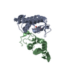
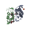
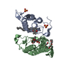
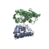
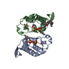
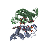
| Title | Crystal structure of bothropstoxin-I chemically modified by p-bromophenacyl bromide (BPB) - monomeric form at a high resolution | |||||||||
 Components Components | Phospholipase A2 homolog bothropstoxin-1 | |||||||||
 Keywords Keywords |  TOXIN / Lys49-PLA2s / Phospholipase homologue / bothropstoxin-I / p-bromophenacyl bromide / myotoxicity / Antibiotic / Antimicrobial / Disulfide bond / Myotoxin / Secreted TOXIN / Lys49-PLA2s / Phospholipase homologue / bothropstoxin-I / p-bromophenacyl bromide / myotoxicity / Antibiotic / Antimicrobial / Disulfide bond / Myotoxin / Secreted | |||||||||
| Function / homology |  Function and homology informationphospholipase A2 activity / arachidonic acid secretion / phospholipid metabolic process / lipid catabolic process / heparin binding / toxin activity / defense response to bacterium / calcium ion binding / extracellular region Function and homology informationphospholipase A2 activity / arachidonic acid secretion / phospholipid metabolic process / lipid catabolic process / heparin binding / toxin activity / defense response to bacterium / calcium ion binding / extracellular regionSimilarity search - Function | |||||||||
| Biological species |  Bothrops jararacussu (jararacussu) Bothrops jararacussu (jararacussu) | |||||||||
| Method | X-RAY DIFFRACTION / SYNCHROTRON / MOLECULAR REPLACEMENT / Resolution: 1.48 Å | |||||||||
 Authors Authors | Marchi-Salvador, D.P. / Fernandes, C.A.H. / Soares, A.M. / Fontes, M.R.M. | |||||||||
 Citation Citation | Journal: J.Struct.Biol. / Year: 2010 Title: Comparison between apo and complexed structures of bothropstoxin-I reveals the role of Lys122 and Ca(2+)-binding loop region for the catalytically inactive Lys49-PLA(2)s. Authors: Fernandes, C.A. / Marchi-Salvador, D.P. / Salvador, G.M. / Silva, M.C. / Costa, T.R. / Soares, A.M. / Fontes, M.R. | |||||||||
| History |
|
- Structure visualization
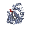
| Structure viewer | Molecule: MolmilJmol/JSmol |
|---|
- Downloads & links
-Download
| PDBx/mmCIF format | 3i03.cif.gz | 48.5 KB | Display | PDBx/mmCIF format |
|---|---|---|---|---|
| PDB format | pdb3i03.ent.gz | 33.5 KB | Display | PDB format |
| PDBx/mmJSON format | 3i03.json.gz | Tree view | PDBx/mmJSON format | |
| Others |  Other downloads Other downloads |
-Validation report
| Arichive directory | https://data.pdbj.org/pub/pdb/validation_reports/i0/3i03ftp://data.pdbj.org/pub/pdb/validation_reports/i0/3i03 | HTTPS FTP |
|---|
-Related structure data
| Related structure data |  3hzdC 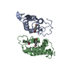 3hzwSC 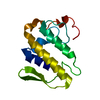 3i3iC  3iq3C S: Starting model for refinement C: citing same article ( |
|---|---|
| Similar structure data |


-Links
 PDBj
PDBj

- Assembly
| Deposited unit | 
| ||||||||
|---|---|---|---|---|---|---|---|---|---|
| 1 | 
| ||||||||
| Unit cell |
|
-Components
| #1: Protein | Mass: 13753.136 Da / Num. of mol.: 1 / Source method: isolated from a natural source Details: The bothropstoxin-I was isolated from Bothrops jararacussu venom. Source: (natural) Bothrops jararacussu (jararacussu) / Tissue: venom / References: UniProt: Q90249 |
|---|---|
| #2: Chemical | ChemComp-PBP /   Mass: 277.941 Da / Num. of mol.: 1 / Source method: obtained synthetically / Formula: C8H6Br2O Mass: 277.941 Da / Num. of mol.: 1 / Source method: obtained synthetically / Formula: C8H6Br2O |
| #3: Chemical | ChemComp-IPA / Isopropyl alcohol  Mass: 60.095 Da / Num. of mol.: 1 / Source method: obtained synthetically / Formula: C3H8O / Comment: alkaloid*YM Mass: 60.095 Da / Num. of mol.: 1 / Source method: obtained synthetically / Formula: C3H8O / Comment: alkaloid*YM |
| #4: Water | ChemComp-HOH / Water Mass: 18.015 Da / Num. of mol.: 351 / Source method: isolated from a natural source / Formula: H2O Mass: 18.015 Da / Num. of mol.: 351 / Source method: isolated from a natural source / Formula: H2O |
-Experimental details
-Experiment
| Experiment | Method: X-RAY DIFFRACTION / Number of used crystals: 1 |
|---|
- Sample preparation
| Crystal | Density Matthews: 2.66 Å3/Da / Density % sol: 53.7 % |
|---|---|
| Crystal grow | Temperature: 283 K / Method: vapor diffusion, hanging drop / pH: 5.8 Details: 20% w/v PEG 4000, 20% v/v Isopropanol, 0.1 M Sodium citrate pH 6.0, VAPOR DIFFUSION, HANGING DROP, temperature 283K |
-Data collection
| Diffraction | Mean temperature: 100 K |
|---|---|
| Diffraction source | Source: SYNCHROTRON / Site: LNLS  / Beamline: W01B-MX2 / Wavelength: 1.425 Å / Beamline: W01B-MX2 / Wavelength: 1.425 Å |
| Detector | Type: MARMOSAIC 225 mm CCD / Detector: CCD / Date: Apr 18, 2007 |
| Radiation | Monochromator: Si(111) double crystal / Protocol: SINGLE WAVELENGTH / Monochromatic (M) / Laue (L): M / Scattering type: x-ray |
| Radiation wavelength | Wavelength: 1.425 Å / Relative weight: 1 |
| Reflection | Resolution: 1.48→50 Å / Num. all: 22361 / Num. obs: 22198 / % possible obs: 99.27 % / Observed criterion σ(I): 1 / Biso Wilson estimate: 24.8 Å2 |
| Reflection shell | Resolution: 1.48→1.53 Å |
- Processing
| Software |
| ||||||||||||||||||||||||||||||||||||||||||||||||||||||||||||||||||||||||||||||||||||||||||||||||||||||||||||||||||||||||||||||||||||||||||||||||||||||||||||||||||||||||||
|---|---|---|---|---|---|---|---|---|---|---|---|---|---|---|---|---|---|---|---|---|---|---|---|---|---|---|---|---|---|---|---|---|---|---|---|---|---|---|---|---|---|---|---|---|---|---|---|---|---|---|---|---|---|---|---|---|---|---|---|---|---|---|---|---|---|---|---|---|---|---|---|---|---|---|---|---|---|---|---|---|---|---|---|---|---|---|---|---|---|---|---|---|---|---|---|---|---|---|---|---|---|---|---|---|---|---|---|---|---|---|---|---|---|---|---|---|---|---|---|---|---|---|---|---|---|---|---|---|---|---|---|---|---|---|---|---|---|---|---|---|---|---|---|---|---|---|---|---|---|---|---|---|---|---|---|---|---|---|---|---|---|---|---|---|---|---|---|---|---|---|---|
| Refinement | Method to determine structure: MOLECULAR REPLACEMENT Starting model: PDB entry 3HZW Resolution: 1.48→45.98 Å / Cor.coef. Fo:Fc: 0.96 / Cor.coef. Fo:Fc free: 0.952 / SU B: 1.304 / SU ML: 0.051 / Isotropic thermal model: OVERALL / Cross valid method: THROUGHOUT / ESU R: 0.095 / ESU R Free: 0.088 / Stereochemistry target values: MAXIMUM LIKELIHOOD / Details: HYDROGENS HAVE BEEN ADDED IN THE RIDING POSITIONS
| ||||||||||||||||||||||||||||||||||||||||||||||||||||||||||||||||||||||||||||||||||||||||||||||||||||||||||||||||||||||||||||||||||||||||||||||||||||||||||||||||||||||||||
| Solvent computation | Ion probe radii: 0.8 Å / Shrinkage radii: 0.8 Å / VDW probe radii: 1.2 Å / Solvent model: MASK | ||||||||||||||||||||||||||||||||||||||||||||||||||||||||||||||||||||||||||||||||||||||||||||||||||||||||||||||||||||||||||||||||||||||||||||||||||||||||||||||||||||||||||
| Displacement parameters | Biso mean: 27.884 Å2
| ||||||||||||||||||||||||||||||||||||||||||||||||||||||||||||||||||||||||||||||||||||||||||||||||||||||||||||||||||||||||||||||||||||||||||||||||||||||||||||||||||||||||||
| Refine analyze |
| ||||||||||||||||||||||||||||||||||||||||||||||||||||||||||||||||||||||||||||||||||||||||||||||||||||||||||||||||||||||||||||||||||||||||||||||||||||||||||||||||||||||||||
| Refinement step | Cycle: LAST / Resolution: 1.48→45.98 Å
| ||||||||||||||||||||||||||||||||||||||||||||||||||||||||||||||||||||||||||||||||||||||||||||||||||||||||||||||||||||||||||||||||||||||||||||||||||||||||||||||||||||||||||
| Refine LS restraints |
| ||||||||||||||||||||||||||||||||||||||||||||||||||||||||||||||||||||||||||||||||||||||||||||||||||||||||||||||||||||||||||||||||||||||||||||||||||||||||||||||||||||||||||
| LS refinement shell | Resolution: 1.48→1.518 Å / Total num. of bins used: 20
| ||||||||||||||||||||||||||||||||||||||||||||||||||||||||||||||||||||||||||||||||||||||||||||||||||||||||||||||||||||||||||||||||||||||||||||||||||||||||||||||||||||||||||
| Xplor file |
|
