 Movie
Movie Controller

[English] 日本語
 Yorodumi
- PDB-4kf3: Crystal Structure of Myotoxin II (MjTX-II), a myotoxic Lys49-phos... -
+ Open data

- Basic information
| Entry | Database: PDB / ID: 4kf3 | ||||||
|---|---|---|---|---|---|---|---|
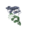
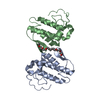
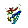
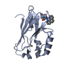
| Title | Crystal Structure of Myotoxin II (MjTX-II), a myotoxic Lys49-phospholipase A2 from Bothrops moojeni. | ||||||
 Components Components | Basic phospholipase A2 homolog 2 | ||||||
 Keywords Keywords |  TOXIN / Phospholipase A2-like myotoxin / Venom glands TOXIN / Phospholipase A2-like myotoxin / Venom glands | ||||||
| Function / homology |  Function and homology information Function and homology informationcalcium-dependent phospholipase A2 activity / arachidonic acid secretion / defense response to fungus / phospholipid metabolic process / lipid catabolic process / negative regulation of T cell proliferation / phospholipid binding / toxin activity / killing of cells of another organism / defense response to bacterium ...calcium-dependent phospholipase A2 activity / arachidonic acid secretion / defense response to fungus / phospholipid metabolic process / lipid catabolic process / negative regulation of T cell proliferation / phospholipid binding / toxin activity / killing of cells of another organism / defense response to bacterium / calcium ion binding / extracellular regionSimilarity search - Function | ||||||
| Biological species |  Bothrops moojeni (Brazilian lancehead) Bothrops moojeni (Brazilian lancehead) | ||||||
| Method | X-RAY DIFFRACTION / SYNCHROTRON / MOLECULAR REPLACEMENT / Resolution: 1.92 Å | ||||||
 Authors Authors | Salvador, G.H.M. / dos Santos, J.I. / Fontes, M.R.M. | ||||||
 Citation Citation | Journal: Toxicon / Year: 2013 Title: Structural and functional studies with mytoxin II from Bothrops moojeni reveal remarkable similarities and differences compared to other catalytically inactive phospholipases A2-like. Authors: Salvador, G.H. / Cavalcante, W.L. / Dos Santos, J.I. / Gallacci, M. / Soares, A.M. / Fontes, M.R. | ||||||
| History |
|
- Structure visualization
| Structure viewer | Molecule: MolmilJmol/JSmol |
|---|
- Downloads & links
-Download
| PDBx/mmCIF format | 4kf3.cif.gz | 67.1 KB | Display | PDBx/mmCIF format |
|---|---|---|---|---|
| PDB format | pdb4kf3.ent.gz | 49.1 KB | Display | PDB format |
| PDBx/mmJSON format | 4kf3.json.gz | Tree view | PDBx/mmJSON format | |
| Others |  Other downloads Other downloads |
-Validation report
| Arichive directory | https://data.pdbj.org/pub/pdb/validation_reports/kf/4kf3ftp://data.pdbj.org/pub/pdb/validation_reports/kf/4kf3 | HTTPS FTP |
|---|
-Related structure data
| Related structure data | 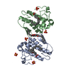 1xxsS S: Starting model for refinement |
|---|---|
| Similar structure data |


-Links
 PDBj
PDBj

- Assembly
| Deposited unit | 
| ||||||||
|---|---|---|---|---|---|---|---|---|---|
| 1 | 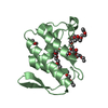
| ||||||||
| 2 | 
| ||||||||
| Unit cell |
|
-Components
| #1: Protein | Mass: 13912.211 Da / Num. of mol.: 2 / Fragment: Phospholipase A2-Like myotoxin / Source method: isolated from a natural source / Source: (natural) Bothrops moojeni (Brazilian lancehead) / References: UniProt: Q9I834#2: Chemical | ChemComp-PE4 / Polyethylene glycol  Mass: 354.436 Da / Num. of mol.: 4 / Source method: obtained synthetically / Formula: C16H34O8 / Comment: precipitant*YM Mass: 354.436 Da / Num. of mol.: 4 / Source method: obtained synthetically / Formula: C16H34O8 / Comment: precipitant*YM#3: Chemical | ChemComp-IPA / Isopropyl alcohol  Mass: 60.095 Da / Num. of mol.: 6 / Source method: obtained synthetically / Formula: C3H8O / Comment: alkaloid*YM Mass: 60.095 Da / Num. of mol.: 6 / Source method: obtained synthetically / Formula: C3H8O / Comment: alkaloid*YM#4: Water | ChemComp-HOH / | Water Mass: 18.015 Da / Num. of mol.: 185 / Source method: isolated from a natural source / Formula: H2O Mass: 18.015 Da / Num. of mol.: 185 / Source method: isolated from a natural source / Formula: H2O |
|---|
-Experimental details
-Experiment
| Experiment | Method: X-RAY DIFFRACTION / Number of used crystals: 1 |
|---|
- Sample preparation
| Crystal | Density Matthews: 2.4 Å3/Da / Density % sol: 48.79 % |
|---|---|
| Crystal grow | Temperature: 291 K / Method: vapor diffusion, hanging drop / pH: 5.6 Details: 20% (v/v) 2-propanol, 20% (w/v) polyethylene Glycol 4000 and 0.1 M Sodium Citrate, pH 5.6, VAPOR DIFFUSION, HANGING DROP, temperature 291K |
-Data collection
| Diffraction | Mean temperature: 100 K |
|---|---|
| Diffraction source | Source: SYNCHROTRON / Site: LNLS  / Beamline: W01B-MX2 / Wavelength: 1.42 Å / Beamline: W01B-MX2 / Wavelength: 1.42 Å |
| Detector | Type: MARMOSAIC 225 mm CCD / Detector: CCD / Date: Mar 22, 2012 / Details: mirrors |
| Radiation | Monochromator: Si(111) double-crystal / Protocol: SINGLE WAVELENGTH / Monochromatic (M) / Laue (L): M / Scattering type: x-ray |
| Radiation wavelength | Wavelength: 1.42 Å / Relative weight: 1 |
| Reflection | Resolution: 1.92→50 Å / Num. obs: 19571 / % possible obs: 92.4 % / Observed criterion σ(F): 0 / Observed criterion σ(I): -3 / Redundancy: 3.9 % / Biso Wilson estimate: 24.6 Å2 / Rmerge(I) obs: 0.044 / Net I/σ(I): 10.37 |
| Reflection shell | Resolution: 1.92→1.99 Å / Redundancy: 3.7 % / Rmerge(I) obs: 0.41 / Mean I/σ(I) obs: 2.01 / % possible all: 97.6 |
- Processing
| Software |
| |||||||||||||||||||||||||
|---|---|---|---|---|---|---|---|---|---|---|---|---|---|---|---|---|---|---|---|---|---|---|---|---|---|---|
| Refinement | Method to determine structure: MOLECULAR REPLACEMENT Starting model: PDB ENTRY 1XXS Resolution: 1.92→38.96 Å / Cross valid method: THROUGHOUT / σ(F): 0 / Stereochemistry target values: Engh & Huber
| |||||||||||||||||||||||||
| Displacement parameters | Biso mean: 56.13 Å2
| |||||||||||||||||||||||||
| Refine analyze | Luzzati coordinate error obs: 0.27 Å / Luzzati d res low obs: 5 Å / Luzzati sigma a obs: 0.23 Å | |||||||||||||||||||||||||
| Refinement step | Cycle: LAST / Resolution: 1.92→38.96 Å
| |||||||||||||||||||||||||
| Refine LS restraints |
|
