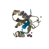
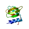
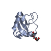
 Movie
Movie Controller


+ Open data

- Basic information
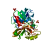
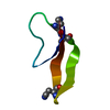
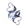
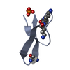
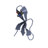
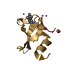
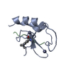
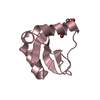
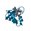
| Entry | Database: PDB / ID: 3hj2 | ||||||
|---|---|---|---|---|---|---|---|
| Title | Crystal structure of covalent dimer of HNP1 | ||||||
 Components Components | HUMAN NEUTROPHIL PEPTIDE 1 | ||||||
 Keywords Keywords |  ANTIMICROBIAL PROTEIN / defensin / antimicrobial / chemotactic / cationic / Cys-rich / covalent dimer / synthetic / Antibiotic / Antiviral defense / Disulfide bond / Fungicide / Phosphoprotein / Secreted ANTIMICROBIAL PROTEIN / defensin / antimicrobial / chemotactic / cationic / Cys-rich / covalent dimer / synthetic / Antibiotic / Antiviral defense / Disulfide bond / Fungicide / Phosphoprotein / Secreted | ||||||
| Function / homology |  Function and homology information Function and homology informationpore-forming activity / Defensins / disruption of plasma membrane integrity in another organism / killing by host of symbiont cells / T cell chemotaxis / Alpha-defensins / defense response to protozoan / intracellular estrogen receptor signaling pathway / defense response to fungus / innate immune response in mucosa ...pore-forming activity / Defensins / disruption of plasma membrane integrity in another organism / killing by host of symbiont cells / T cell chemotaxis / Alpha-defensins / defense response to protozoan / intracellular estrogen receptor signaling pathway / defense response to fungus / innate immune response in mucosa / Golgi lumen / chemotaxis / azurophil granule lumen / antimicrobial humoral immune response mediated by antimicrobial peptide / antibacterial humoral response / collagen-containing extracellular matrix / defense response to virus / killing of cells of another organism / cellular response to lipopolysaccharide / defense response to Gram-negative bacterium / defense response to Gram-positive bacterium / immune response / Neutrophil degranulation / extracellular space / extracellular exosome / extracellular regionSimilarity search - Function | ||||||
| Biological species |  Homo sapiens (human) Homo sapiens (human) | ||||||
| Method | X-RAY DIFFRACTION / SYNCHROTRON / MOLECULAR REPLACEMENT / Resolution: 1.4 Å | ||||||
 Authors Authors | Lubkowski, J. / Pazgier, M. / Lu, W. | ||||||
 Citation Citation | Journal: To be Published Title: What Dictates the Multifaced Functions of the Human alpha-Defensin HNP1? Authors: Wei, G. / de Leeuw, E. / Pazgier, M. / Rajabi, M. / Li, J. / Zou, G. / Ericksen, B. / Wu, Z. / Yuan, W. / Szmacinski, H. / Lu, W.-Y. / Lubkowski, J. / Lehrer, R.L. / Lu, W. | ||||||
| History |
|
- Structure visualization
| Structure viewer | Molecule: MolmilJmol/JSmol |
|---|
- Downloads & links
-Download
| PDBx/mmCIF format | 3hj2.cif.gz | 37.9 KB | Display | PDBx/mmCIF format |
|---|---|---|---|---|
| PDB format | pdb3hj2.ent.gz | 27.2 KB | Display | PDB format |
| PDBx/mmJSON format | 3hj2.json.gz | Tree view | PDBx/mmJSON format | |
| Others |  Other downloads Other downloads |
-Validation report
| Arichive directory | https://data.pdbj.org/pub/pdb/validation_reports/hj/3hj2ftp://data.pdbj.org/pub/pdb/validation_reports/hj/3hj2 | HTTPS FTP |
|---|
-Related structure data
| Related structure data |  1dfnS S: Starting model for refinement |
|---|---|
| Similar structure data |


-Links
 PDBj
PDBj


- Assembly
| Deposited unit | 
| ||||||||
|---|---|---|---|---|---|---|---|---|---|
| 1 |
| ||||||||
| Unit cell |
|
-Components
| #1: Protein/peptide | Mass: 3669.358 Da / Num. of mol.: 2 / Fragment: Residues 65-94 / Source method: obtained synthetically / Source: (synth.) Homo sapiens (human) / References: UniProt: P59665#2: Water | ChemComp-HOH / | Water Mass: 18.015 Da / Num. of mol.: 51 / Source method: isolated from a natural source / Formula: H2O Mass: 18.015 Da / Num. of mol.: 51 / Source method: isolated from a natural source / Formula: H2OSequence details | AN ARTIFICIAL | |
|---|
-Experimental details
-Experiment
| Experiment | Method: X-RAY DIFFRACTION / Number of used crystals: 1 |
|---|
- Sample preparation
| Crystal | Density Matthews: 1.98 Å3/Da / Density % sol: 37.83 % |
|---|---|
| Crystal grow | Temperature: 293 K / Method: vapor diffusion, hanging drop / pH: 5.6 Details: 0.1 M Na-citrate (pH 5.6), 0.2 M NH4-acetate, 30% (v/v) MPD, VAPOR DIFFUSION, HANGING DROP, temperature 293.0K |
-Data collection
| Diffraction | Mean temperature: 100 K |
|---|---|
| Diffraction source | Source: SYNCHROTRON / Site: APS  / Beamline: 22-ID / Wavelength: 1 Å / Beamline: 22-ID / Wavelength: 1 Å |
| Detector | Type: MARMOSAIC 300 mm CCD / Detector: CCD / Date: Aug 10, 2007 Details: Rosenbaum-Rock monochromator high-resolution double-crystal Si(220) |
| Radiation | Monochromator: Si crystal / Protocol: SINGLE WAVELENGTH / Monochromatic (M) / Laue (L): M / Scattering type: x-ray |
| Radiation wavelength | Wavelength: 1 Å / Relative weight: 1 |
| Reflection | Resolution: 1.4→24.68 Å / Num. all: 11611 / Num. obs: 11611 / % possible obs: 96.3 % / Observed criterion σ(F): -1.7 / Observed criterion σ(I): -3 / Redundancy: 6.2 % / Biso Wilson estimate: 22.2 Å2 / Rmerge(I) obs: 0.065 / Rsym value: 0.065 / Net I/σ(I): 22.5 |
| Reflection shell | Resolution: 1.4→1.45 Å / Redundancy: 6.4 % / Rmerge(I) obs: 0.114 / Mean I/σ(I) obs: 17.8 / Num. unique all: 1171 / Rsym value: 0.114 / % possible all: 100 |
- Processing
| Software |
| ||||||||||||||||||||||||||||||||||||||||||||||||||||||||||||||||||||||||||||||||
|---|---|---|---|---|---|---|---|---|---|---|---|---|---|---|---|---|---|---|---|---|---|---|---|---|---|---|---|---|---|---|---|---|---|---|---|---|---|---|---|---|---|---|---|---|---|---|---|---|---|---|---|---|---|---|---|---|---|---|---|---|---|---|---|---|---|---|---|---|---|---|---|---|---|---|---|---|---|---|---|---|---|
| Refinement | Method to determine structure: MOLECULAR REPLACEMENT Starting model: 1dfn Resolution: 1.4→15 Å / Cor.coef. Fo:Fc: 0.939 / Cor.coef. Fo:Fc free: 0.922 / SU B: 2.655 / SU ML: 0.047 / Cross valid method: THROUGHOUT / ESU R: 0.085 / ESU R Free: 0.075 / Stereochemistry target values: MAXIMUM LIKELIHOOD / Details: HYDROGENS HAVE BEEN ADDED IN THE RIDING POSITIONS
| ||||||||||||||||||||||||||||||||||||||||||||||||||||||||||||||||||||||||||||||||
| Solvent computation | Ion probe radii: 0.8 Å / Shrinkage radii: 0.8 Å / VDW probe radii: 1.2 Å / Solvent model: BABINET MODEL WITH MASK | ||||||||||||||||||||||||||||||||||||||||||||||||||||||||||||||||||||||||||||||||
| Displacement parameters | Biso mean: 25.827 Å2
| ||||||||||||||||||||||||||||||||||||||||||||||||||||||||||||||||||||||||||||||||
| Refine analyze | Luzzati coordinate error obs: 0.248 Å / Luzzati d res low obs: 15 Å | ||||||||||||||||||||||||||||||||||||||||||||||||||||||||||||||||||||||||||||||||
| Refinement step | Cycle: LAST / Resolution: 1.4→15 Å
| ||||||||||||||||||||||||||||||||||||||||||||||||||||||||||||||||||||||||||||||||
| Refine LS restraints |
| ||||||||||||||||||||||||||||||||||||||||||||||||||||||||||||||||||||||||||||||||
| LS refinement shell | Resolution: 1.4→1.436 Å / Total num. of bins used: 20
| ||||||||||||||||||||||||||||||||||||||||||||||||||||||||||||||||||||||||||||||||
| Refinement TLS params. | Method: refined / Refine-ID: X-RAY DIFFRACTION
| ||||||||||||||||||||||||||||||||||||||||||||||||||||||||||||||||||||||||||||||||
| Refinement TLS group |
|
