 Movie
Movie Controller


+ Open data

- Basic information
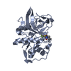
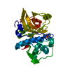
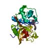
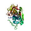
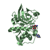
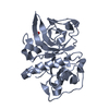
| Entry | Database: PDB / ID: 4bpv | ||||||
|---|---|---|---|---|---|---|---|
| Title | MOUSE CATHEPSIN S WITH COVALENT LIGAND | ||||||
 Components Components | CATHEPSIN S | ||||||
 Keywords Keywords | HYDROLASE / CYSTEINE PROTEASE / COVALENT LIGAND | ||||||
| Function / homology |  Function and homology information Function and homology informationTrafficking and processing of endosomal TLR / Assembly of collagen fibrils and other multimeric structures / cathepsin S / Degradation of the extracellular matrix / basement membrane disassembly / positive regulation of cation channel activity / antigen processing and presentation of peptide antigen / MHC class II antigen presentation / response to acidic pH / proteoglycan binding ...Trafficking and processing of endosomal TLR / Assembly of collagen fibrils and other multimeric structures / cathepsin S / Degradation of the extracellular matrix / basement membrane disassembly / positive regulation of cation channel activity / antigen processing and presentation of peptide antigen / MHC class II antigen presentation / response to acidic pH / proteoglycan binding / cysteine-type endopeptidase activator activity involved in apoptotic process / fibronectin binding / collagen catabolic process / laminin binding / bone resorption / cysteine-type peptidase activity / phagocytic vesicle / collagen binding / Neutrophil degranulation / proteolysis involved in protein catabolic process / early endosome lumen / positive regulation of apoptotic signaling pathway / protein processing / positive regulation of inflammatory response / antigen processing and presentation of exogenous peptide antigen via MHC class II / late endosome / peptidase activity / lysosome / immune response / cysteine-type endopeptidase activity / intracellular membrane-bounded organelle / cell surface / proteolysis / extracellular space / membraneSimilarity search - Function | ||||||
| Biological species |  MUS MUSCULUS (house mouse) MUS MUSCULUS (house mouse) | ||||||
| Method | X-RAY DIFFRACTION / SYNCHROTRON / MOLECULAR REPLACEMENT / Resolution: 2 Å | ||||||
 Authors Authors | Banner, D.W. / Benz, J. / Gsell, B. / Stihle, M. / Ruf, A. / Haap, W. | ||||||
 Citation Citation | Journal: To be Published Title: Cathepsin S Nitrile Inhibitors Authors: Haap, W. / Banner, D.W. | ||||||
| History |
|
- Structure visualization
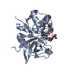
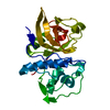
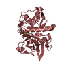
| Structure viewer | Molecule: MolmilJmol/JSmol |
|---|
- Downloads & links
-Download
| PDBx/mmCIF format | 4bpv.cif.gz | 259.9 KB | Display | PDBx/mmCIF format |
|---|---|---|---|---|
| PDB format | pdb4bpv.ent.gz | 211.2 KB | Display | PDB format |
| PDBx/mmJSON format | 4bpv.json.gz | Tree view | PDBx/mmJSON format | |
| Others |  Other downloads Other downloads |
-Validation report
| Arichive directory | https://data.pdbj.org/pub/pdb/validation_reports/bp/4bpvftp://data.pdbj.org/pub/pdb/validation_reports/bp/4bpv | HTTPS FTP |
|---|
-Related structure data


-Links
 PDBj
PDBj


- Assembly
| Deposited unit | 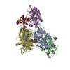
| ||||||||
|---|---|---|---|---|---|---|---|---|---|
| 1 | 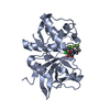
| ||||||||
| 2 | 
| ||||||||
| 3 | 
| ||||||||
| 4 | 
| ||||||||
| 5 | 
| ||||||||
| 6 | 
| ||||||||
| Unit cell |
|
-Components
| #1: Protein | Mass: 24662.480 Da / Num. of mol.: 6 / Fragment: RESIDUES 116-340 Source method: isolated from a genetically manipulated source Source: (gene. exp.) MUS MUSCULUS (house mouse) / Production host:  ESCHERICHIA COLI (E. coli) / References: UniProt: O70370, cathepsin S ESCHERICHIA COLI (E. coli) / References: UniProt: O70370, cathepsin S#2: Chemical | ChemComp-OFH / (   Mass: 603.962 Da / Num. of mol.: 5 / Source method: obtained synthetically / Formula: C23H24ClF6N3O5S Mass: 603.962 Da / Num. of mol.: 5 / Source method: obtained synthetically / Formula: C23H24ClF6N3O5S#3: Water | ChemComp-HOH / | Water Mass: 18.015 Da / Num. of mol.: 938 / Source method: isolated from a natural source / Formula: H2O Mass: 18.015 Da / Num. of mol.: 938 / Source method: isolated from a natural source / Formula: H2OSequence details | 116-340 NO MUTATIONS | |
|---|
-Experimental details
-Experiment
| Experiment | Method: X-RAY DIFFRACTION / Number of used crystals: 1 |
|---|
- Sample preparation
| Crystal | Density Matthews: 3.45 Å3/Da / Density % sol: 64.4 % / Description: NONE |
|---|---|
| Crystal grow | pH: 7.5 / Details: 25% PEG 3350, 0.1 M HEPES PH7.5, 0.2M NH4AC |
-Data collection
| Diffraction | Mean temperature: 100 K |
|---|---|
| Diffraction source | Source: SYNCHROTRON / Site: SLS  / Beamline: X10SA / Wavelength: 1 / Beamline: X10SA / Wavelength: 1 |
| Detector | Type: MARRESEARCH / Detector: CCD / Date: Aug 12, 2009 |
| Radiation | Protocol: SINGLE WAVELENGTH / Monochromatic (M) / Laue (L): M / Scattering type: x-ray |
| Radiation wavelength | Wavelength: 1 Å / Relative weight: 1 |
| Reflection | Resolution: 1.95→45.1 Å / Num. obs: 128672 / % possible obs: 99.6 % / Observed criterion σ(I): -3 / Redundancy: 3.78 % / Rmerge(I) obs: 0.08 / Net I/σ(I): 10.33 |
| Reflection shell | Resolution: 1.95→2 Å / Redundancy: 3.52 % / Rmerge(I) obs: 0.52 / Mean I/σ(I) obs: 1.84 / % possible all: 97.3 |
- Processing
| Software |
| ||||||||||||||||||||||||||||||||||||||||||||||||||||||||||||||||||||||||||||||||||||||||||||||||||||||||||||||||||||||||||||||||||||||||||||||||||||||||||||||||||||||||||||||||||||||
|---|---|---|---|---|---|---|---|---|---|---|---|---|---|---|---|---|---|---|---|---|---|---|---|---|---|---|---|---|---|---|---|---|---|---|---|---|---|---|---|---|---|---|---|---|---|---|---|---|---|---|---|---|---|---|---|---|---|---|---|---|---|---|---|---|---|---|---|---|---|---|---|---|---|---|---|---|---|---|---|---|---|---|---|---|---|---|---|---|---|---|---|---|---|---|---|---|---|---|---|---|---|---|---|---|---|---|---|---|---|---|---|---|---|---|---|---|---|---|---|---|---|---|---|---|---|---|---|---|---|---|---|---|---|---|---|---|---|---|---|---|---|---|---|---|---|---|---|---|---|---|---|---|---|---|---|---|---|---|---|---|---|---|---|---|---|---|---|---|---|---|---|---|---|---|---|---|---|---|---|---|---|---|---|
| Refinement | Method to determine structure: MOLECULAR REPLACEMENT Starting model: IN HOUSE STRUCTURES Resolution: 2→45.11 Å / Cor.coef. Fo:Fc: 0.935 / Cor.coef. Fo:Fc free: 0.906 / SU B: 4.487 / SU ML: 0.123 / Cross valid method: THROUGHOUT / ESU R: 0.167 / ESU R Free: 0.159 / Stereochemistry target values: MAXIMUM LIKELIHOOD Details: HYDROGENS HAVE BEEN ADDED IN THE RIDING POSITIONS BUT NOT OUTPUT. U VALUES REFINED INDIVIDUALLY. CHAINS A,B,C,D FORM LAYERS. E BRIDGES THE LAYERS AND K IS ONLY PARTIALLY VISIBLE
| ||||||||||||||||||||||||||||||||||||||||||||||||||||||||||||||||||||||||||||||||||||||||||||||||||||||||||||||||||||||||||||||||||||||||||||||||||||||||||||||||||||||||||||||||||||||
| Solvent computation | Ion probe radii: 0.8 Å / Shrinkage radii: 0.8 Å / VDW probe radii: 1.2 Å / Solvent model: BABINET MODEL WITH MASK | ||||||||||||||||||||||||||||||||||||||||||||||||||||||||||||||||||||||||||||||||||||||||||||||||||||||||||||||||||||||||||||||||||||||||||||||||||||||||||||||||||||||||||||||||||||||
| Displacement parameters | Biso mean: 33.644 Å2
| ||||||||||||||||||||||||||||||||||||||||||||||||||||||||||||||||||||||||||||||||||||||||||||||||||||||||||||||||||||||||||||||||||||||||||||||||||||||||||||||||||||||||||||||||||||||
| Refinement step | Cycle: LAST / Resolution: 2→45.11 Å
| ||||||||||||||||||||||||||||||||||||||||||||||||||||||||||||||||||||||||||||||||||||||||||||||||||||||||||||||||||||||||||||||||||||||||||||||||||||||||||||||||||||||||||||||||||||||
| Refine LS restraints |
|