 Movie
Movie Controller


+ Open data

- Basic information
| Entry | Database: PDB / ID: 3h4r | ||||||
|---|---|---|---|---|---|---|---|
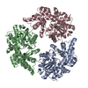
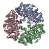
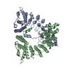
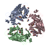
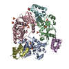
| Title | Crystal structure of E. coli RecE exonuclease | ||||||
 Components Components | Exodeoxyribonuclease 8 | ||||||
 Keywords Keywords | HYDROLASE / Exonuclease / Recombination / Nuclease | ||||||
| Function / homology |  Function and homology information Function and homology informationdouble-stranded DNA 5'-3' DNA exonuclease activity / Hydrolases; Acting on ester bonds; Exodeoxyribonucleases producing 5'-phosphomonoesters / identical protein bindingSimilarity search - Function | ||||||
| Biological species |  Escherichia coli (E. coli) Escherichia coli (E. coli) | ||||||
| Method | X-RAY DIFFRACTION / SYNCHROTRON / SAD / Resolution: 2.8 Å | ||||||
 Authors Authors | Bell, C.E. / Zhang, J. | ||||||
 Citation Citation | Journal: Structure / Year: 2009 Title: Crystal structure of E. coli RecE protein reveals a toroidal tetramer for processing double-stranded DNA breaks. Authors: Zhang, J. / Xing, X. / Herr, A.B. / Bell, C.E. | ||||||
| History |
|
- Structure visualization
| Structure viewer | Molecule: MolmilJmol/JSmol |
|---|
- Downloads & links
-Download
| PDBx/mmCIF format | 3h4r.cif.gz | 56.5 KB | Display | PDBx/mmCIF format |
|---|---|---|---|---|
| PDB format | pdb3h4r.ent.gz | 40.8 KB | Display | PDB format |
| PDBx/mmJSON format | 3h4r.json.gz | Tree view | PDBx/mmJSON format | |
| Others |  Other downloads Other downloads |
-Validation report
| Arichive directory | https://data.pdbj.org/pub/pdb/validation_reports/h4/3h4rftp://data.pdbj.org/pub/pdb/validation_reports/h4/3h4r | HTTPS FTP |
|---|
-Related structure data
| Similar structure data |
|---|


-Links
 PDBj
PDBj- Assembly
| Deposited unit | 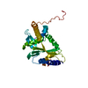
| ||||||||
|---|---|---|---|---|---|---|---|---|---|
| 1 | 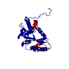
| ||||||||
| Unit cell |
|
-Components
| #1: Protein | / Exodeoxyribonuclease VIII / EXO VIII Mass: 30403.252 Da / Num. of mol.: 1 / Fragment: C-terminal domain: UNP residues 606-866 / Mutation: P658L Source method: isolated from a genetically manipulated source Source: (gene. exp.) Escherichia coli (E. coli) / Strain: K-12 / Gene: b1350, JW1344, recE / Plasmid: pET14b / Production host: Escherichia coli (E. coli) / Strain (production host): BL21(AI)References: UniProt: P15032, Hydrolases; Acting on ester bonds; Exodeoxyribonucleases producing 5'-phosphomonoesters |
|---|
-Experimental details
-Experiment
| Experiment | Method: X-RAY DIFFRACTION / Number of used crystals: 1 |
|---|
- Sample preparation
| Crystal | Density Matthews: 4.2 Å3/Da / Density % sol: 70.7 % |
|---|---|
| Crystal grow | Temperature: 298 K / Method: vapor diffusion, hanging drop / pH: 7 Details: 30-42% Glycerol, 100 mM DL-malic acid, pH 7.0, VAPOR DIFFUSION, HANGING DROP, temperature 298K |
-Data collection
| Diffraction | Mean temperature: 100 K |
|---|---|
| Diffraction source | Source: SYNCHROTRON / Site: APS  / Beamline: 31-ID / Wavelength: 0.97929 Å / Beamline: 31-ID / Wavelength: 0.97929 Å |
| Detector | Type: MAR CCD 165 mm / Detector: CCD / Date: Dec 9, 2007 |
| Radiation | Monochromator: Kohzu HLD-4 Diamond(111) Double Crystal / Protocol: SINGLE WAVELENGTH / Monochromatic (M) / Laue (L): M / Scattering type: x-ray |
| Radiation wavelength | Wavelength: 0.97929 Å / Relative weight: 1 |
| Reflection | Resolution: 2.8→50 Å / Num. all: 13172 / Num. obs: 13172 / % possible obs: 99.4 % / Redundancy: 23.5 % / Biso Wilson estimate: 33.1 Å2 / Rmerge(I) obs: 0.08 / Net I/σ(I): 19.6 |
| Reflection shell | Resolution: 2.8→2.95 Å / Redundancy: 23.4 % / Rmerge(I) obs: 0.7 / Mean I/σ(I) obs: 3.3 / % possible all: 100 |
- Processing
| Software |
| ||||||||||||||||||||||||||||||||||||
|---|---|---|---|---|---|---|---|---|---|---|---|---|---|---|---|---|---|---|---|---|---|---|---|---|---|---|---|---|---|---|---|---|---|---|---|---|---|
| Refinement | Method to determine structure: SAD / Resolution: 2.8→45.43 Å / Rfactor Rfree error: 0.012 / Data cutoff high absF: 3811437.52 / Data cutoff low absF: 0 / Isotropic thermal model: RESTRAINED / Cross valid method: THROUGHOUT / σ(F): 2 / Stereochemistry target values: Engh & Huber / Details: BULK SOLVENT MODEL USED
| ||||||||||||||||||||||||||||||||||||
| Solvent computation | Solvent model: FLAT MODEL / Bsol: 65.5675 Å2 / ksol: 0.35 e/Å3 | ||||||||||||||||||||||||||||||||||||
| Displacement parameters | Biso mean: 97.3 Å2
| ||||||||||||||||||||||||||||||||||||
| Refine analyze |
| ||||||||||||||||||||||||||||||||||||
| Refinement step | Cycle: LAST / Resolution: 2.8→45.43 Å
| ||||||||||||||||||||||||||||||||||||
| Refine LS restraints |
| ||||||||||||||||||||||||||||||||||||
| LS refinement shell | Resolution: 2.8→2.98 Å / Rfactor Rfree error: 0.045 / Total num. of bins used: 6
| ||||||||||||||||||||||||||||||||||||
| Xplor file | Serial no: 1 / Param file: protein_rep.param / Topol file: protein.top |
