 Movie
Movie Controller


+ Open data

- Basic information
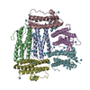
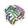
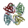
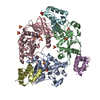
| Entry | Database: PDB / ID: 2f7n | ||||||
|---|---|---|---|---|---|---|---|
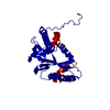
| Title | Structure of D. radiodurans Dps-1 | ||||||
 Components Components | DNA-binding stress response protein, Dps family | ||||||
 Keywords Keywords |  DNA BINDING PROTEIN / 4-helix bundle DNA BINDING PROTEIN / 4-helix bundle | ||||||
| Function / homology |  Function and homology informationOxidoreductases; Oxidizing metal ions / oxidoreductase activity, acting on metal ions / nucleoid / ferric iron binding / intracellular iron ion homeostasis / DNA binding / cytoplasm Function and homology informationOxidoreductases; Oxidizing metal ions / oxidoreductase activity, acting on metal ions / nucleoid / ferric iron binding / intracellular iron ion homeostasis / DNA binding / cytoplasmSimilarity search - Function | ||||||
| Biological species |  Deinococcus radiodurans (radioresistant) Deinococcus radiodurans (radioresistant) | ||||||
| Method | X-RAY DIFFRACTION / SAD / Resolution: 2 Å | ||||||
 Authors Authors | Lee, Y.H. / Kim, S.G. / Bhattacharyya, G. / Grove, A. | ||||||
 Citation Citation | Journal: J.Mol.Biol. / Year: 2006 Title: Crystal structure of Dps-1, a functionally distinct Dps protein from Deinococcus radiodurans. Authors: Kim, S.G. / Bhattacharyya, G. / Grove, A. / Lee, Y.H. | ||||||
| History |
|
- Structure visualization
| Structure viewer | Molecule: MolmilJmol/JSmol |
|---|
- Downloads & links
-Download
| PDBx/mmCIF format | 2f7n.cif.gz | 51.4 KB | Display | PDBx/mmCIF format |
|---|---|---|---|---|
| PDB format | pdb2f7n.ent.gz | 36.5 KB | Display | PDB format |
| PDBx/mmJSON format | 2f7n.json.gz | Tree view | PDBx/mmJSON format | |
| Others |  Other downloads Other downloads |
-Validation report
| Arichive directory | https://data.pdbj.org/pub/pdb/validation_reports/f7/2f7nftp://data.pdbj.org/pub/pdb/validation_reports/f7/2f7n | HTTPS FTP |
|---|
-Related structure data
| Similar structure data |
|---|


-Links
 PDBj
PDBj


- Assembly
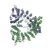
| Deposited unit | 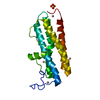
| ||||||||||||||||||||||||
|---|---|---|---|---|---|---|---|---|---|---|---|---|---|---|---|---|---|---|---|---|---|---|---|---|---|
| 1 | x 6
| ||||||||||||||||||||||||
| 2 | x 12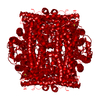
| ||||||||||||||||||||||||
| Unit cell |
| ||||||||||||||||||||||||
| Components on special symmetry positions |
|
-Components
| #1: Protein | Mass: 23050.719 Da / Num. of mol.: 1 Source method: isolated from a genetically manipulated source Source: (gene. exp.) Deinococcus radiodurans (radioresistant)Plasmid: pET3 / Species (production host): Escherichia coli / Production host: Escherichia coli BL21(DE3) (bacteria) / Strain (production host): BL21DE3 / References: GenBank: 15807254, UniProt: Q9RS64*PLUS | ||||
|---|---|---|---|---|---|
| #2: Chemical | ChemComp-CO /   Mass: 58.933 Da / Num. of mol.: 4 / Source method: obtained synthetically / Formula: Co Mass: 58.933 Da / Num. of mol.: 4 / Source method: obtained synthetically / Formula: Co#3: Chemical | ChemComp-SO4 / | Sulfate  Mass: 96.063 Da / Num. of mol.: 1 / Source method: obtained synthetically / Formula: SO4 Mass: 96.063 Da / Num. of mol.: 1 / Source method: obtained synthetically / Formula: SO4#4: Water | ChemComp-HOH / | Water Mass: 18.015 Da / Num. of mol.: 115 / Source method: isolated from a natural source / Formula: H2O Mass: 18.015 Da / Num. of mol.: 115 / Source method: isolated from a natural source / Formula: H2O |
-Experimental details
-Experiment
| Experiment | Method: X-RAY DIFFRACTION / Number of used crystals: 1 |
|---|
- Sample preparation
| Crystal | Density Matthews: 2.65 Å3/Da / Density % sol: 53.52 % |
|---|---|
| Crystal grow | Temperature: 293 K / Method: vapor diffusion, sitting drop / pH: 6.5 Details: 100mM MES; 100mM cobalt chloride; 2M anmmonium sulfate, pH 6.5, VAPOR DIFFUSION, SITTING DROP, temperature 293K |
-Data collection
| Diffraction | Mean temperature: 100 K |
|---|---|
| Diffraction source | Source: ROTATING ANODE / Type: RIGAKU RU200 / Wavelength: 1.5418 Å |
| Detector | Type: MARRESEARCH / Detector: IMAGE PLATE / Date: May 14, 2005 |
| Radiation | Monochromator: silicon / Protocol: SINGLE WAVELENGTH / Monochromatic (M) / Laue (L): M / Scattering type: x-ray |
| Radiation wavelength | Wavelength: 1.5418 Å / Relative weight: 1 |
| Reflection | Resolution: 2→30 Å / Num. all: 31997 / Num. obs: 16568 / % possible obs: 99.8 % / Observed criterion σ(I): 5 / Redundancy: 9.2 % / Rmerge(I) obs: 0.11 / Net I/σ(I): 14.7 |
| Reflection shell | Resolution: 2→2.09 Å / Redundancy: 8.4 % / Rmerge(I) obs: 0.48 / Mean I/σ(I) obs: 1.9 / % possible all: 90.3 |
- Processing
| Software |
| ||||||||||||||||||||||||||||||||
|---|---|---|---|---|---|---|---|---|---|---|---|---|---|---|---|---|---|---|---|---|---|---|---|---|---|---|---|---|---|---|---|---|---|
| Refinement | Method to determine structure: SAD / Resolution: 2→30 Å / Cross valid method: THROUGHOUT / σ(F): 0 / Stereochemistry target values: Engh & Huber
| ||||||||||||||||||||||||||||||||
| Refine analyze | Luzzati coordinate error obs: 0.22 Å / Luzzati sigma a obs: 0.2 Å | ||||||||||||||||||||||||||||||||
| Refinement step | Cycle: LAST / Resolution: 2→30 Å
| ||||||||||||||||||||||||||||||||
| Refine LS restraints |
|
