[English] 日本語
Yorodumi
- PDB-3gq9: Crystal Structure of the Bacteriophage phi29 gene product 12 N-te... -

+
Open data


ID or keywords:

Loading...

-
Basic information

Entry
Database: PDB / ID: 3gq9
Title
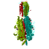
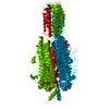
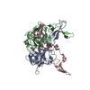
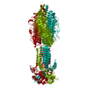
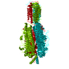
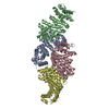
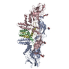
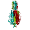
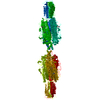
Crystal Structure of the Bacteriophage phi29 gene product 12 N-terminal fragment in an apo form
ComponentsPreneck appendage protein
KeywordsVIRAL PROTEIN / beta helix
Function / homology
Function and homology information


virus tail, fiber / symbiont entry into host cell via disruption of host cell envelope / adhesion receptor-mediated virion attachment to host cell / symbiont entry into host cell / virion attachment to host cell / ATP binding / metal ion binding
Similarity search - Function
Insect antifreeze protein / Peptidase G2, IMC autoproteolytic cleavage domain / Peptidase_G2, IMC autoproteolytic cleavage domain / Pectate lyase superfamily protein / Pectate lyase superfamily protein / Parallel beta-helix repeat / Parallel beta-helix repeats / Single-stranded right-handed beta-helix, Pectin lyase-like / Pectate Lyase C-like / UDP N-Acetylglucosamine Acyltransferase; domain 1 ...Insect antifreeze protein / Peptidase G2, IMC autoproteolytic cleavage domain / Peptidase_G2, IMC autoproteolytic cleavage domain / Pectate lyase superfamily protein / Pectate lyase superfamily protein / Parallel beta-helix repeat / Parallel beta-helix repeats / Single-stranded right-handed beta-helix, Pectin lyase-like / Pectate Lyase C-like / UDP N-Acetylglucosamine Acyltransferase; domain 1 / Pectin lyase fold / Pectin lyase fold/virulence factor / 3 Solenoid / Mainly Beta
Similarity search - Domain/homology
Pre-neck appendage protein / Pre-neck appendage protein
Similarity search - Component
Biological speciesBacillus phage phi29 (virus)
MethodX-RAY DIFFRACTION / SYNCHROTRON / Resolution: 2 Å
AuthorsXiang, Y. / Rossmann, M.G.
CitationJournal: Mol.Cell / Year: 2009
Title: Crystallographic insights into the autocatalytic assembly mechanism of a bacteriophage tail spike.
Authors: Xiang, Y. / Leiman, P.G. / Li, L. / Grimes, S. / Anderson, D.L. / Rossmann, M.G.
History
DepositionMar 24, 2009Deposition site: RCSB / Processing site: RCSB
Revision 1.0May 26, 2009Provider: repository / Type: Initial release
Revision 1.1Jul 13, 2011Group: Version format compliance
Revision 1.2Mar 21, 2012Group: Database references
Revision 1.3Nov 1, 2017Group: Refinement description / Category: software
Revision 1.4Feb 21, 2024Group: Data collection / Database references / Derived calculations
Category: chem_comp_atom / chem_comp_bond ...chem_comp_atom / chem_comp_bond / database_2 / pdbx_struct_conn_angle / pdbx_struct_special_symmetry / struct_conn / struct_ref_seq_dif / struct_site
Item: _database_2.pdbx_DOI / _database_2.pdbx_database_accession ..._database_2.pdbx_DOI / _database_2.pdbx_database_accession / _pdbx_struct_conn_angle.ptnr1_auth_comp_id / _pdbx_struct_conn_angle.ptnr1_auth_seq_id / _pdbx_struct_conn_angle.ptnr1_label_asym_id / _pdbx_struct_conn_angle.ptnr1_label_atom_id / _pdbx_struct_conn_angle.ptnr1_label_comp_id / _pdbx_struct_conn_angle.ptnr1_label_seq_id / _pdbx_struct_conn_angle.ptnr3_auth_comp_id / _pdbx_struct_conn_angle.ptnr3_auth_seq_id / _pdbx_struct_conn_angle.ptnr3_label_asym_id / _pdbx_struct_conn_angle.ptnr3_label_atom_id / _pdbx_struct_conn_angle.ptnr3_label_comp_id / _pdbx_struct_conn_angle.ptnr3_label_seq_id / _pdbx_struct_conn_angle.value / _struct_conn.pdbx_dist_value / _struct_conn.ptnr1_auth_comp_id / _struct_conn.ptnr1_auth_seq_id / _struct_conn.ptnr1_label_asym_id / _struct_conn.ptnr1_label_atom_id / _struct_conn.ptnr1_label_comp_id / _struct_conn.ptnr1_label_seq_id / _struct_conn.ptnr2_auth_comp_id / _struct_conn.ptnr2_auth_seq_id / _struct_conn.ptnr2_label_asym_id / _struct_conn.ptnr2_label_atom_id / _struct_conn.ptnr2_label_comp_id / _struct_conn.ptnr2_label_seq_id / _struct_ref_seq_dif.details / _struct_site.pdbx_auth_asym_id / _struct_site.pdbx_auth_comp_id / _struct_site.pdbx_auth_seq_id

-
Structure visualization

Structure viewerMolecule:
MolmilJmol/JSmol

Downloads & links

-
Assembly

Deposited unit
A: Preneck appendage protein
hetero molecules


Theoretical massNumber of molelcules
Total (without water)64,6644
Polymers64,4111
Non-polymers2533
Water6,539363
1
A: Preneck appendage protein
hetero molecules

A: Preneck appendage protein
hetero molecules

A: Preneck appendage protein
hetero molecules


Theoretical massNumber of molelcules
Total (without water)193,99312
Polymers193,2333
Non-polymers7609
Water543
TypeNameSymmetry operationNumber
identity operation1_555x,y,z1
crystal symmetry operation2_655-y+1,x-y,z1
crystal symmetry operation3_665-x+y+1,-x+1,z1
Buried area27760 Å2
ΔGint-202 kcal/mol
Surface area47380 Å2
MethodPISA
2
A: Preneck appendage protein
hetero molecules
x 6


Theoretical massNumber of molelcules
Total (without water)387,98524
Polymers386,4656
Non-polymers1,52018
Water1086
TypeNameSymmetry operationNumber
identity operation1_555x,y,z1
crystal symmetry operation2_655-y+1,x-y,z1
crystal symmetry operation3_665-x+y+1,-x+1,z1
crystal symmetry operation16_545y+1/3,x-1/3,-z+2/31
crystal symmetry operation17_555x-y+1/3,-y+2/3,-z+2/31
crystal symmetry operation18_655-x+4/3,-x+y+2/3,-z+2/31
Buried area56780 Å2
ΔGint-420 kcal/mol
Surface area93510 Å2
MethodPISA
Unit cell
Length a, b, c (Å)89.633, 89.633, 585.719
Angle α, β, γ (deg.)90.000, 90.000, 120.000
Int Tables number155
Space group name H-MH32
Components on special symmetry positions
IDModelComponents
11A-858-

HOH

-
Components

#1: Protein Preneck appendage protein


Mass: 64410.914 Da / Num. of mol.: 1 / Fragment: D1*D2D3, Residues 89-691
Source method: isolated from a genetically manipulated source
Details: RESIDUES 89-854 EXPRESSED WITH A N-TERMIINAL HIS6 TAG
Source: (gene. exp.) Bacillus phage phi29 (virus) / Gene: 12, gene product 12 / Plasmid: PET28B / Production host: Escherichia coli (E. coli) / Strain (production host): BL21(DE3)RIL / References: UniProt: B3VMP8, UniProt: P20345*PLUS
#2: Chemical ChemComp-NA / SODIUM ION


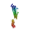
Mass: 22.990 Da / Num. of mol.: 2 / Source method: obtained synthetically / Formula: Na
#3: Chemical ChemComp-NHE / 2-[N-CYCLOHEXYLAMINO]ETHANE SULFONIC ACID / N-CYCLOHEXYLTAURINE / CHES / CHES (buffer)


Mass: 207.290 Da / Num. of mol.: 1 / Source method: obtained synthetically / Formula: C8H17NO3S / Comment: pH buffer*YM
#4: Water ChemComp-HOH / water / Water


Mass: 18.015 Da / Num. of mol.: 363 / Source method: isolated from a natural source / Formula: H2O
Sequence detailsTHESE MUTATIONS MIGHT BE AN ACCUMULATED RESULT OF THE PHAGE PASSAGING

-
Experimental details

-
Experiment

ExperimentMethod: X-RAY DIFFRACTION / Number of used crystals: 1

-
Sample preparation

CrystalDensity Matthews: 3.51 Å3/Da / Density % sol: 65.01 %
Crystal growTemperature: 298 K / Method: vapor diffusion, hanging drop / pH: 10
Details: 100mM CHES at ~pH 10.0 and 8% PEG3K. Crystalls obtained were then soaked in a mother liquid containing 10mM EDTA for ~5 hours., VAPOR DIFFUSION, HANGING DROP, temperature 298K

-
Data collection

DiffractionMean temperature: 100 K
Diffraction sourceSource: SYNCHROTRON / Site: APS / Beamline: 14-ID-B / Wavelength: 0.99 Å
DetectorType: MAR CCD 165 mm / Detector: CCD / Date: Apr 21, 2008
RadiationMonochromator: SI(111) double-crystal / Protocol: SINGLE WAVELENGTH / Monochromatic (M) / Laue (L): M / Scattering type: x-ray
Radiation wavelengthWavelength: 0.99 Å / Relative weight: 1
ReflectionResolution: 2→43.69 Å / Num. obs: 61778 / % possible obs: 99.4 % / Redundancy: 7 % / Rmerge(I) obs: 0.079 / Χ2: 0.944 / Net I/σ(I): 24.302
Reflection shell
Resolution (Å)Redundancy (%)Rmerge(I) obsNum. unique allΧ2Diffraction-ID% possible all
2-2.075.90.39760461.186198.7
2.07-2.156.10.31160600.893199
2.15-2.256.40.24260720.924199.3
2.25-2.376.80.20160980.825199.3
2.37-2.527.20.16661350.8199.5
2.52-2.717.60.12361640.834199.6
2.71-2.997.70.09361740.886199.7
2.99-3.427.70.06462030.951199.9
3.42-4.317.50.04563101.087199.9
4.31-5070.03965161.081199.5

-
Processing

Software
NameVersionClassificationNB
DENZOdata reduction
SCALEPACKdata scaling
REFMAC5.2.0019refinement
PDB_EXTRACT3.006data extraction
HKL-2000data reduction
HKL-2000data scaling
RefinementResolution: 2→43.69 Å / Cor.coef. Fo:Fc: 0.962 / Cor.coef. Fo:Fc free: 0.95 / Occupancy max: 1 / Occupancy min: 0.33 / SU B: 2.745 / SU ML: 0.076 / Cross valid method: THROUGHOUT / σ(F): 0 / ESU R: 0.127 / ESU R Free: 0.122 / Stereochemistry target values: MAXIMUM LIKELIHOOD
RfactorNum. reflection% reflectionSelection details
Rfree0.204 3077 5 %RANDOM
Rwork0.172 ---
obs0.174 61145 98.47 %-
Solvent computationIon probe radii: 0.8 Å / Shrinkage radii: 0.8 Å / VDW probe radii: 1.2 Å / Solvent model: BABINET MODEL WITH MASK
Displacement parametersBiso max: 57.16 Å2 / Biso mean: 23.137 Å2 / Biso min: 10.84 Å2
Baniso -1Baniso -2Baniso -3
1-0.28 Å20.14 Å20 Å2
2--0.28 Å20 Å2
3----0.41 Å2
Refinement stepCycle: LAST / Resolution: 2→43.69 Å
ProteinNucleic acidLigandSolventTotal
Num. atoms4478 0 15 363 4856
Refine LS restraints
Refine-IDTypeDev idealDev ideal targetNumber
X-RAY DIFFRACTIONr_bond_refined_d0.010.0214655
X-RAY DIFFRACTIONr_angle_refined_deg1.2621.9416338
X-RAY DIFFRACTIONr_dihedral_angle_1_deg6.7755634
X-RAY DIFFRACTIONr_dihedral_angle_2_deg39.27324.439205
X-RAY DIFFRACTIONr_dihedral_angle_3_deg12.08815739
X-RAY DIFFRACTIONr_dihedral_angle_4_deg15.7191528
X-RAY DIFFRACTIONr_chiral_restr0.0890.2721
X-RAY DIFFRACTIONr_gen_planes_refined0.0040.023586
X-RAY DIFFRACTIONr_nbd_refined0.2050.22190
X-RAY DIFFRACTIONr_nbtor_refined0.3020.23275
X-RAY DIFFRACTIONr_xyhbond_nbd_refined0.1160.2361
X-RAY DIFFRACTIONr_metal_ion_refined0.1370.25
X-RAY DIFFRACTIONr_symmetry_vdw_refined0.1720.2172
X-RAY DIFFRACTIONr_symmetry_hbond_refined0.1230.242
X-RAY DIFFRACTIONr_mcbond_it0.4931.53011
X-RAY DIFFRACTIONr_mcangle_it0.96724851
X-RAY DIFFRACTIONr_scbond_it1.81631681
X-RAY DIFFRACTIONr_scangle_it2.9754.51471
LS refinement shellResolution: 2→2.054 Å / Total num. of bins used: 20
RfactorNum. reflection% reflection
Rfree0.272 212 -
Rwork0.22 4149 -
all-4361 -
obs--97.11 %

+
About Yorodumi

-
News

-
Feb 9, 2022. New format data for meta-information of EMDB entries

New format data for meta-information of EMDB entries

  • Version 3 of the EMDB header file is now the official format.
  • The previous official version 1.9 will be removed from the archive.

Related info.:EMDB header

External links:wwPDB to switch to version 3 of the EMDB data model

-
Aug 12, 2020. Covid-19 info

Covid-19 info

URL: https://pdbj.org/emnavi/covid19.php

New page: Covid-19 featured information page in EM Navigator.

Related info.:Covid-19 info / Mar 5, 2020. Novel coronavirus structure data

+
Mar 5, 2020. Novel coronavirus structure data

Novel coronavirus structure data

Related info.:Yorodumi Speices / Aug 12, 2020. Covid-19 info

External links:COVID-19 featured content - PDBj / Molecule of the Month (242):Coronavirus Proteases

+
Jan 31, 2019. EMDB accession codes are about to change! (news from PDBe EMDB page)

EMDB accession codes are about to change! (news from PDBe EMDB page)

  • The allocation of 4 digits for EMDB accession codes will soon come to an end. Whilst these codes will remain in use, new EMDB accession codes will include an additional digit and will expand incrementally as the available range of codes is exhausted. The current 4-digit format prefixed with “EMD-” (i.e. EMD-XXXX) will advance to a 5-digit format (i.e. EMD-XXXXX), and so on. It is currently estimated that the 4-digit codes will be depleted around Spring 2019, at which point the 5-digit format will come into force.
  • The EM Navigator/Yorodumi systems omit the EMD- prefix.

Related info.:Q: What is EMD? / ID/Accession-code notation in Yorodumi/EM Navigator

External links:EMDB Accession Codes are Changing Soon! / Contact to PDBj

+
Jul 12, 2017. Major update of PDB

Major update of PDB

  • wwPDB released updated PDB data conforming to the new PDBx/mmCIF dictionary.
  • This is a major update changing the version number from 4 to 5, and with Remediation, in which all the entries are updated.
  • In this update, many items about electron microscopy experimental information are reorganized (e.g. em_software).
  • Now, EM Navigator and Yorodumi are based on the updated data.

External links:wwPDB Remediation / Enriched Model Files Conforming to OneDep Data Standards Now Available in the PDB FTP Archive

-
Yorodumi

Thousand views of thousand structures

  • Yorodumi is a browser for structure data from EMDB, PDB, SASBDB, etc.
  • This page is also the successor to EM Navigator detail page, and also detail information page/front-end page for Omokage search.
  • The word "yorodu" (or yorozu) is an old Japanese word meaning "ten thousand". "mi" (miru) is to see.

Related info.:EMDB / PDB / SASBDB / Comparison of 3 databanks / Yorodumi Search / Aug 31, 2016. New EM Navigator & Yorodumi / Yorodumi Papers / Jmol/JSmol / Function and homology information / Changes in new EM Navigator and Yorodumi

Read more