 Movie
Movie Controller


+ Open data

- Basic information
| Entry | Database: PDB / ID: 3j2p | |||||||||
|---|---|---|---|---|---|---|---|---|---|---|
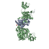
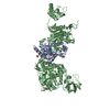
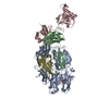
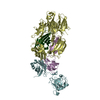
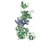
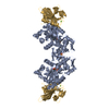
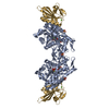
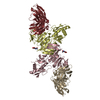
| Title | CryoEM structure of Dengue virus envelope protein heterotetramer | |||||||||
 Components Components |
| |||||||||
 Keywords Keywords | VIRAL PROTEIN / flavivirus / fusion protein / protein complex / membrane | |||||||||
| Function / homology |  Function and homology information Function and homology informationsymbiont-mediated suppression of host JAK-STAT cascade via inhibition of host TYK2 activity / flavivirin / host cell mitochondrion / symbiont-mediated suppression of host JAK-STAT cascade via inhibition of STAT2 activity / symbiont-mediated suppression of host cytoplasmic pattern recognition receptor signaling pathway via inhibition of MAVS activity / viral capsid / double-stranded RNA binding / nucleoside-triphosphate phosphatase / protein complex oligomerization / monoatomic ion channel activity ...symbiont-mediated suppression of host JAK-STAT cascade via inhibition of host TYK2 activity / flavivirin / host cell mitochondrion / symbiont-mediated suppression of host JAK-STAT cascade via inhibition of STAT2 activity / symbiont-mediated suppression of host cytoplasmic pattern recognition receptor signaling pathway via inhibition of MAVS activity / viral capsid / double-stranded RNA binding / nucleoside-triphosphate phosphatase / protein complex oligomerization / monoatomic ion channel activity / mRNA (guanine-N7)-methyltransferase / methyltransferase cap1 / clathrin-dependent endocytosis of virus by host cell / mRNA (nucleoside-2'-O-)-methyltransferase activity / mRNA 5'-cap (guanine-N7-)-methyltransferase activity / RNA helicase activity / host cell perinuclear region of cytoplasm / host cell endoplasmic reticulum membrane / protein dimerization activity / symbiont-mediated suppression of host type I interferon-mediated signaling pathway / RNA helicase / induction by virus of host autophagy / serine-type endopeptidase activity / RNA-directed RNA polymerase / viral RNA genome replication / virus-mediated perturbation of host defense response / RNA-dependent RNA polymerase activity / fusion of virus membrane with host endosome membrane / viral envelope / host cell nucleus / virion attachment to host cell / structural molecule activity / virion membrane / ATP hydrolysis activity / proteolysis / extracellular region / ATP binding / membrane / metal ion binding Similarity search - Function | |||||||||
| Biological species |  Dengue virus 2 Dengue virus 2 | |||||||||
| Method | ELECTRON MICROSCOPY / single particle reconstruction / cryo EM / Resolution: 3.6 Å | |||||||||
 Authors Authors | Zhang, X. / Ge, P. / Yu, X. / Brannan, J.M. / Bi, G. / Zhang, Q. / Schein, S. / Zhou, Z.H. | |||||||||
 Citation Citation | Journal: Nat Struct Mol Biol / Year: 2013 Title: Cryo-EM structure of the mature dengue virus at 3.5-Å resolution. Authors: Xiaokang Zhang / Peng Ge / Xuekui Yu / Jennifer M Brannan / Guoqiang Bi / Qinfen Zhang / Stan Schein / Z Hong Zhou /  Abstract: Regulated by pH, membrane-anchored proteins E and M function during dengue virus maturation and membrane fusion. Our atomic model of the whole virion from cryo-electron microscopy at 3.5-Å ...Regulated by pH, membrane-anchored proteins E and M function during dengue virus maturation and membrane fusion. Our atomic model of the whole virion from cryo-electron microscopy at 3.5-Å resolution reveals that in the mature virus at neutral extracellular pH, the N-terminal 20-amino-acid segment of M (involving three pH-sensing histidines) latches and thereby prevents spring-loaded E fusion protein from prematurely exposing its fusion peptide. This M latch is fastened at an earlier stage, during maturation at acidic pH in the trans-Golgi network. At a later stage, to initiate infection in response to acidic pH in the late endosome, M releases the latch and exposes the fusion peptide. Thus, M serves as a multistep chaperone of E to control the conformational changes accompanying maturation and infection. These pH-sensitive interactions could serve as targets for drug discovery. | |||||||||
| History |
|
- Structure visualization
| Movie |
Movie viewer |
|---|---|
| Structure viewer | Molecule: MolmilJmol/JSmol |
- Downloads & links
-Download
| PDBx/mmCIF format | 3j2p.cif.gz | 228.5 KB | Display | PDBx/mmCIF format |
|---|---|---|---|---|
| PDB format | pdb3j2p.ent.gz | 188.6 KB | Display | PDB format |
| PDBx/mmJSON format | 3j2p.json.gz | Tree view | PDBx/mmJSON format | |
| Others |  Other downloads Other downloads |
-Validation report
| Summary document | 3j2p_validation.pdf.gz | 1.1 MB | Display | wwPDB validaton report |
|---|---|---|---|---|
| Full document | 3j2p_full_validation.pdf.gz | 1.2 MB | Display | |
| Data in XML | 3j2p_validation.xml.gz | 58.1 KB | Display | |
| Data in CIF | 3j2p_validation.cif.gz | 83 KB | Display | |
| Arichive directory | https://data.pdbj.org/pub/pdb/validation_reports/j2/3j2pftp://data.pdbj.org/pub/pdb/validation_reports/j2/3j2p | HTTPS FTP |
-Related structure data
| Related structure data | 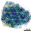 5499MC 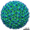 5520C  3j27C M: map data used to model this data C: citing same article ( |
|---|---|
| Similar structure data |


-Links
 PDBj
PDBj


- Assembly
| Deposited unit | 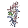
|
|---|---|
| 1 |
|
-Components
| #1: Protein | Mass: 54363.734 Da / Num. of mol.: 2 / Fragment: UNP residues 281-775 / Source method: isolated from a natural source / Source: (natural) Dengue virus 2 / Cell line: Mosquito Cells C6/36 / Strain: New Guinea / References: UniProt: P14340#2: Protein | Mass: 8259.646 Da / Num. of mol.: 2 / Fragment: UNP residues 206-280 / Source method: isolated from a natural source / Source: (natural) Dengue virus 2 / Cell line: Mosquito Cells C6/36 / Strain: New Guinea / References: UniProt: P14340#3: Polysaccharide | Source method: isolated from a genetically manipulated source #4: Sugar |   Type: D-saccharide, beta linking / Mass: 221.208 Da / Num. of mol.: 2 Type: D-saccharide, beta linking / Mass: 221.208 Da / Num. of mol.: 2Source method: isolated from a genetically manipulated source Formula: C8H15NO6 Sequence details | THE AUTHORS STATE THAT R15A (UNP R220A) IN SMALL ENVELOPE PROTEIN M IS CORRECT FOR THE NEW GUINEA STRAIN. | |
|---|
-Experimental details
-Experiment
| Experiment | Method: ELECTRON MICROSCOPY |
|---|---|
| EM experiment | Aggregation state: PARTICLE / 3D reconstruction method: single particle reconstruction |
- Sample preparation
| Component | Name: Dengue Virus 2 / Type: VIRUS Details: E:M:M:E heterotetramer. icosahedral virion with envelope |
|---|---|
| Molecular weight | Value: 0.125 MDa / Experimental value: NO |
| Details of virus | Empty: NO / Enveloped: YES / Host category: VERTEBRATES / Isolate: STRAIN / Type: VIRION |
| Natural host | Organism: Homo sapiens |
| Buffer solution | Name: TNE 50 mM Tris, 140 mM NaCl, 5 mM EDTA / pH: 7.4 / Details: TNE 50 mM Tris, 140 mM NaCl, 5 mM EDTA |
| Specimen | Embedding applied: NO / Shadowing applied: NO / Staining applied: NO / Vitrification applied: YES / Details: 50 mM Tris, 140 mM NaCl, 5 mM EDTA |
| Specimen support | Details: Quantifoil R2/1 |
| Vitrification | Instrument: HOMEMADE PLUNGER / Cryogen name: ETHANE Details: 2.5 uL sample added per grid, plunged into liquid ethane Method: 2.5 uL sample added per grid |
- Electron microscopy imaging
| Experimental equipment |  Model: Titan Krios / Image courtesy: FEI Company |
|---|---|
| Microscopy | Model: FEI TITAN KRIOS / Date: Dec 23, 2010 |
| Electron gun | Electron source:  FIELD EMISSION GUN / Accelerating voltage: 300 kV / Illumination mode: FLOOD BEAM FIELD EMISSION GUN / Accelerating voltage: 300 kV / Illumination mode: FLOOD BEAM |
| Electron lens | Mode: BRIGHT FIELD / Nominal magnification: 59000 X / Calibrated magnification: 57518 X / Nominal defocus max: 2400 nm / Nominal defocus min: 500 nm / Cs: 2.7 mm / Astigmatism: software compensation / Camera length: 0 mm |
| Specimen holder | Specimen holder model: FEI TITAN KRIOS AUTOGRID HOLDER / Tilt angle max: 0 ° / Tilt angle min: 0 ° |
| Image recording | Electron dose: 25 e/Å2 / Film or detector model: KODAK SO-163 FILM / Details: Scanned by Nikon 9000ED |
| Image scans | Num. digital images: 1103 |
- Processing
| EM software | Name: EMAN / Category: 3D reconstruction | ||||||||||||
|---|---|---|---|---|---|---|---|---|---|---|---|---|---|
| CTF correction | Details: EMAN, per particle, with astigmatism compensation | ||||||||||||
| Symmetry | Point symmetry: I (icosahedral) | ||||||||||||
| 3D reconstruction | Method: MPSA / Resolution: 3.6 Å / Resolution method: FSC 0.143 CUT-OFF / Num. of particles: 9288 / Nominal pixel size: 1.076 Å / Actual pixel size: 1.104 Å Magnification calibration: previously calibrated with TMV pitch Details: EMAN with Multi-path Simulated Annealing / Symmetry type: POINT | ||||||||||||
| Refinement step | Cycle: LAST
|
