 Movie
Movie Controller

[English] 日本語
 Yorodumi
- PDB-3fnu: Crystal structure of KNI-10006 bound histo-aspartic protease (HAP... -
+ Open data

- Basic information
| Entry | Database: PDB / ID: 3fnu | ||||||
|---|---|---|---|---|---|---|---|
| Title | Crystal structure of KNI-10006 bound histo-aspartic protease (HAP) from Plasmodium falciparum | ||||||
 Components Components | HAP protein | ||||||
 Keywords Keywords | HYDROLASE/HYDROLASE INHIBITOR / Histo-aspartic protease /  HYDROLASE / Plasmepsin / Aspartic protease / KNI / KNI-10006 / HYDROLASE-HYDROLASE INHIBITOR COMPLEX HYDROLASE / Plasmepsin / Aspartic protease / KNI / KNI-10006 / HYDROLASE-HYDROLASE INHIBITOR COMPLEX | ||||||
| Function / homology |  Function and homology information Function and homology informationMHC class II antigen presentation / plasmepsin II / acquisition of nutrients from host / Neutrophil degranulation / vacuolar lumen / food vacuole / aspartic-type endopeptidase activity / proteolysis / membraneSimilarity search - Function | ||||||
| Biological species |  Plasmodium falciparum (malaria parasite P. falciparum) Plasmodium falciparum (malaria parasite P. falciparum) | ||||||
| Method | X-RAY DIFFRACTION / SYNCHROTRON / MOLECULAR REPLACEMENT / Resolution: 3 Å | ||||||
 Authors Authors | Bhaumik, P. / Gustchina, A. / Wlodawer, A. | ||||||
 Citation Citation | Journal: J.Mol.Biol. / Year: 2009 Title: Crystal structures of the histo-aspartic protease (HAP) from Plasmodium falciparum. Authors: Bhaumik, P. / Xiao, H. / Parr, C.L. / Kiso, Y. / Gustchina, A. / Yada, R.Y. / Wlodawer, A. | ||||||
| History |
|
- Structure visualization
| Structure viewer | Molecule: MolmilJmol/JSmol |
|---|
- Downloads & links
-Download
| PDBx/mmCIF format | 3fnu.cif.gz | 261.4 KB | Display | PDBx/mmCIF format |
|---|---|---|---|---|
| PDB format | pdb3fnu.ent.gz | 220.6 KB | Display | PDB format |
| PDBx/mmJSON format | 3fnu.json.gz | Tree view | PDBx/mmJSON format | |
| Others |  Other downloads Other downloads |
-Validation report
| Arichive directory | https://data.pdbj.org/pub/pdb/validation_reports/fn/3fnuftp://data.pdbj.org/pub/pdb/validation_reports/fn/3fnu | HTTPS FTP |
|---|
-Related structure data


-Links
 PDBj
PDBj- Assembly
| Deposited unit | 
| ||||||||||||||||||||||||||||||||||||||||||||||||
|---|---|---|---|---|---|---|---|---|---|---|---|---|---|---|---|---|---|---|---|---|---|---|---|---|---|---|---|---|---|---|---|---|---|---|---|---|---|---|---|---|---|---|---|---|---|---|---|---|---|
| 1 | 
| ||||||||||||||||||||||||||||||||||||||||||||||||
| 2 | 
| ||||||||||||||||||||||||||||||||||||||||||||||||
| Unit cell |
| ||||||||||||||||||||||||||||||||||||||||||||||||
| Noncrystallographic symmetry (NCS) | NCS domain:
NCS domain segments:
NCS ensembles :
| ||||||||||||||||||||||||||||||||||||||||||||||||
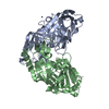
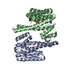
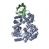
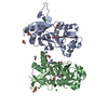
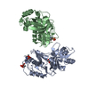
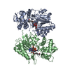
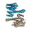
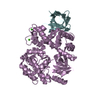
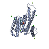
| Details | Four molecules of histo-aspartic protease (HAP) are present in the asymmetric unit. These four molecules are present as two dimers. Each protein molecule is complexed to one KNI-10006 molecule. |
-Components
| #1: Protein | Mass: 37435.250 Da / Num. of mol.: 4 / Fragment: Histo-aspartic protease Source method: isolated from a genetically manipulated source Source: (gene. exp.) Plasmodium falciparum (malaria parasite P. falciparum)Strain: 3D7 / Gene: HAP, PF14_0078 / Plasmid: pET32b(+) / Production host:  Escherichia coli (E. coli) / Strain (production host): Rosetta-gami B (DE3)pLysS / References: UniProt: Q8IM15 Escherichia coli (E. coli) / Strain (production host): Rosetta-gami B (DE3)pLysS / References: UniProt: Q8IM15#2: Chemical | ChemComp-006 / ( 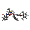  Type: peptide-like, Peptide-like / Class: Inhibitor / Mass: 631.782 Da / Num. of mol.: 4 / Source method: obtained synthetically / Formula: C35H41N3O6S / References: KNI-10006 Type: peptide-like, Peptide-like / Class: Inhibitor / Mass: 631.782 Da / Num. of mol.: 4 / Source method: obtained synthetically / Formula: C35H41N3O6S / References: KNI-10006#3: Water | ChemComp-HOH / | Water Mass: 18.015 Da / Num. of mol.: 95 / Source method: isolated from a natural source / Formula: H2O Mass: 18.015 Da / Num. of mol.: 95 / Source method: isolated from a natural source / Formula: H2O |
|---|
-Experimental details
-Experiment
| Experiment | Method: X-RAY DIFFRACTION / Number of used crystals: 1 |
|---|
- Sample preparation
| Crystal | Density Matthews: 3.19 Å3/Da / Density % sol: 61.42 % Description: AUTHORS STATE THAT THE VALUE OF RMERGE IN THE HIGHEST RESOLUTION SHELL IS HIGH DUE TO POORLY DIFFRACTING CRYSTAL/HIGH SYMMETRY SPACE GROUP/HIGHLY REDUNDANT DATA. CRYSTAL HAS SUFFERED SOME RADIATION DAMAGE |
|---|---|
| Crystal grow | Temperature: 293 K / Method: vapor diffusion, hanging drop Details: 0.2M KH2PO4, 20% PEG 3350, VAPOR DIFFUSION, HANGING DROP, temperature 293K |
-Data collection
| Diffraction | Mean temperature: 100 K |
|---|---|
| Diffraction source | Source: SYNCHROTRON / Site: APS  / Beamline: 22-ID / Wavelength: 0.99999 Å / Beamline: 22-ID / Wavelength: 0.99999 Å |
| Detector | Type: MARMOSAIC 300 mm CCD / Detector: CCD |
| Radiation | Protocol: SINGLE WAVELENGTH / Monochromatic (M) / Laue (L): M / Scattering type: x-ray |
| Radiation wavelength | Wavelength: 0.99999 Å / Relative weight: 1 |
| Reflection | Resolution: 3→40 Å / Num. all: 39071 / Num. obs: 39041 / % possible obs: 99.9 % / Redundancy: 12.3 % / Rmerge(I) obs: 0.128 |
| Reflection shell | Resolution: 3→3.1 Å / Redundancy: 14.1 % / Rmerge(I) obs: 1.642 / Mean I/σ(I) obs: 1.7 / Num. unique all: 3590 / % possible all: 100 |
- Processing
| Software |
| |||||||||||||||||||||||||||||||||||||||||||||||||||||||||||||||||||||||||||||||||||||||||||||||||||||||||||||||||||||||||||||||||||||||||||||||||||||||||||||||||||||||||||||||||||||||||||||||||||||||||||||||||||||||||||||||||
|---|---|---|---|---|---|---|---|---|---|---|---|---|---|---|---|---|---|---|---|---|---|---|---|---|---|---|---|---|---|---|---|---|---|---|---|---|---|---|---|---|---|---|---|---|---|---|---|---|---|---|---|---|---|---|---|---|---|---|---|---|---|---|---|---|---|---|---|---|---|---|---|---|---|---|---|---|---|---|---|---|---|---|---|---|---|---|---|---|---|---|---|---|---|---|---|---|---|---|---|---|---|---|---|---|---|---|---|---|---|---|---|---|---|---|---|---|---|---|---|---|---|---|---|---|---|---|---|---|---|---|---|---|---|---|---|---|---|---|---|---|---|---|---|---|---|---|---|---|---|---|---|---|---|---|---|---|---|---|---|---|---|---|---|---|---|---|---|---|---|---|---|---|---|---|---|---|---|---|---|---|---|---|---|---|---|---|---|---|---|---|---|---|---|---|---|---|---|---|---|---|---|---|---|---|---|---|---|---|---|---|---|---|---|---|---|---|---|---|---|---|---|---|---|---|---|---|
| Refinement | Method to determine structure: MOLECULAR REPLACEMENT / Resolution: 3→30 Å / Cor.coef. Fo:Fc: 0.941 / Cor.coef. Fo:Fc free: 0.932 / SU B: 44.709 / SU ML: 0.38 / TLS residual ADP flag: LIKELY RESIDUAL / Cross valid method: THROUGHOUT / ESU R Free: 0.415 / Stereochemistry target values: MAXIMUM LIKELIHOOD
| |||||||||||||||||||||||||||||||||||||||||||||||||||||||||||||||||||||||||||||||||||||||||||||||||||||||||||||||||||||||||||||||||||||||||||||||||||||||||||||||||||||||||||||||||||||||||||||||||||||||||||||||||||||||||||||||||
| Solvent computation | Ion probe radii: 0.8 Å / Shrinkage radii: 0.8 Å / VDW probe radii: 1.2 Å / Solvent model: MASK | |||||||||||||||||||||||||||||||||||||||||||||||||||||||||||||||||||||||||||||||||||||||||||||||||||||||||||||||||||||||||||||||||||||||||||||||||||||||||||||||||||||||||||||||||||||||||||||||||||||||||||||||||||||||||||||||||
| Displacement parameters | Biso mean: 53.564 Å2
| |||||||||||||||||||||||||||||||||||||||||||||||||||||||||||||||||||||||||||||||||||||||||||||||||||||||||||||||||||||||||||||||||||||||||||||||||||||||||||||||||||||||||||||||||||||||||||||||||||||||||||||||||||||||||||||||||
| Refinement step | Cycle: LAST / Resolution: 3→30 Å
| |||||||||||||||||||||||||||||||||||||||||||||||||||||||||||||||||||||||||||||||||||||||||||||||||||||||||||||||||||||||||||||||||||||||||||||||||||||||||||||||||||||||||||||||||||||||||||||||||||||||||||||||||||||||||||||||||
| Refine LS restraints |
| |||||||||||||||||||||||||||||||||||||||||||||||||||||||||||||||||||||||||||||||||||||||||||||||||||||||||||||||||||||||||||||||||||||||||||||||||||||||||||||||||||||||||||||||||||||||||||||||||||||||||||||||||||||||||||||||||
| Refine LS restraints NCS | Dom-ID: 1 / Number: 2598 / Refine-ID: X-RAY DIFFRACTION
| |||||||||||||||||||||||||||||||||||||||||||||||||||||||||||||||||||||||||||||||||||||||||||||||||||||||||||||||||||||||||||||||||||||||||||||||||||||||||||||||||||||||||||||||||||||||||||||||||||||||||||||||||||||||||||||||||
| LS refinement shell | Resolution: 3→3.077 Å / Total num. of bins used: 20
| |||||||||||||||||||||||||||||||||||||||||||||||||||||||||||||||||||||||||||||||||||||||||||||||||||||||||||||||||||||||||||||||||||||||||||||||||||||||||||||||||||||||||||||||||||||||||||||||||||||||||||||||||||||||||||||||||
| Refinement TLS params. | Method: refined / Refine-ID: X-RAY DIFFRACTION
| |||||||||||||||||||||||||||||||||||||||||||||||||||||||||||||||||||||||||||||||||||||||||||||||||||||||||||||||||||||||||||||||||||||||||||||||||||||||||||||||||||||||||||||||||||||||||||||||||||||||||||||||||||||||||||||||||
| Refinement TLS group |
|
