 Movie
Movie Controller

[English] 日本語
 Yorodumi
- PDB-3fnt: Crystal structure of pepstatin A bound histo-aspartic protease (H... -
+ Open data

- Basic information
| Entry | Database: PDB / ID: 3fnt | ||||||
|---|---|---|---|---|---|---|---|
| Title | Crystal structure of pepstatin A bound histo-aspartic protease (HAP) from Plasmodium falciparum | ||||||
 Components Components |
| ||||||
 Keywords Keywords | HYDROLASE/HYDROLASE INHIBITOR / Histo-aspartic protease / HAP /  Plasmepsin / Pepstatin A / Aspartic protease / HYDROLASE-HYDROLASE INHIBITOR COMPLEX Plasmepsin / Pepstatin A / Aspartic protease / HYDROLASE-HYDROLASE INHIBITOR COMPLEX | ||||||
| Function / homology |  Function and homology information Function and homology informationMHC class II antigen presentation / plasmepsin II / acquisition of nutrients from host / Neutrophil degranulation / vacuolar lumen / food vacuole / aspartic-type endopeptidase activity / proteolysis / membraneSimilarity search - Function | ||||||
| Biological species |  Plasmodium falciparum (malaria parasite P. falciparum) Plasmodium falciparum (malaria parasite P. falciparum) Streptomyces argenteolus subsp. toyonakensis (bacteria) Streptomyces argenteolus subsp. toyonakensis (bacteria) | ||||||
| Method | X-RAY DIFFRACTION / MOLECULAR REPLACEMENT / Resolution: 3.3 Å | ||||||
 Authors Authors | Bhaumik, P. / Gustchina, A. / Wlodawer, A. | ||||||
 Citation Citation | Journal: J.Mol.Biol. / Year: 2009 Title: Crystal structures of the histo-aspartic protease (HAP) from plasmodium falciparum. Authors: Bhaumik, P. / Xiao, H. / Parr, C.L. / Kiso, Y. / Gustchina, A. / Yada, R.Y. / Wlodawer, A. | ||||||
| History |
|
- Structure visualization
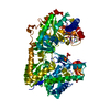
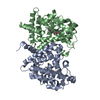
| Structure viewer | Molecule: MolmilJmol/JSmol |
|---|
- Downloads & links
-Download
| PDBx/mmCIF format | 3fnt.cif.gz | 81.1 KB | Display | PDBx/mmCIF format |
|---|---|---|---|---|
| PDB format | pdb3fnt.ent.gz | 59.9 KB | Display | PDB format |
| PDBx/mmJSON format | 3fnt.json.gz | Tree view | PDBx/mmJSON format | |
| Others |  Other downloads Other downloads |
-Validation report
| Arichive directory | https://data.pdbj.org/pub/pdb/validation_reports/fn/3fntftp://data.pdbj.org/pub/pdb/validation_reports/fn/3fnt | HTTPS FTP |
|---|
-Related structure data
| Related structure data | 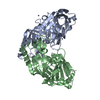 3fnsC 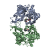 3fnuC  1smeS C: citing same article ( S: Starting model for refinement |
|---|---|
| Similar structure data |


-Links
 PDBj
PDBj
- Assembly
| Deposited unit | 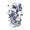
| ||||||||
|---|---|---|---|---|---|---|---|---|---|
| 1 | 
| ||||||||
| 2 |
| ||||||||
| Unit cell |
| ||||||||
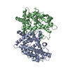
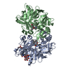
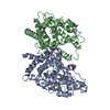
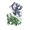
| Details | One molecule of histo-aspartic protease (HAP) is present in the asymmetric unit. One pepstatin A molecule is bound to the active site of the enzyme |
-Components
| #1: Protein | Mass: 37435.250 Da / Num. of mol.: 1 / Fragment: Histo-aspartic protease (HAP) Source method: isolated from a genetically manipulated source Source: (gene. exp.) Plasmodium falciparum (malaria parasite P. falciparum)Strain: 3D7 / Gene: HAP, PF14_0078 / Plasmid: pET32b(+) / Production host: Escherichia coli (E. coli) / Strain (production host): Rosetta-gami B (DE3)pLysS / References: UniProt: Q8IM15 | ||
|---|---|---|---|
| #2: Protein/peptide | Enzyme inhibitor / 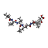  Type: Oligopeptide / Class: Enzyme inhibitor / Mass: 685.891 Da / Num. of mol.: 1 / Source method: obtained synthetically / Details: unspecified Type: Oligopeptide / Class: Enzyme inhibitor / Mass: 685.891 Da / Num. of mol.: 1 / Source method: obtained synthetically / Details: unspecifiedSource: (synth.) Streptomyces argenteolus subsp. toyonakensis (bacteria)References: Pepstatin | ||
| #3: Chemical | Ethylene glycol  Mass: 62.068 Da / Num. of mol.: 2 / Source method: obtained synthetically / Formula: C2H6O2 Mass: 62.068 Da / Num. of mol.: 2 / Source method: obtained synthetically / Formula: C2H6O2#4: Water | ChemComp-HOH / | Water Mass: 18.015 Da / Num. of mol.: 9 / Source method: isolated from a natural source / Formula: H2O Mass: 18.015 Da / Num. of mol.: 9 / Source method: isolated from a natural source / Formula: H2O |
-Experimental details
-Experiment
| Experiment | Method: X-RAY DIFFRACTION / Number of used crystals: 1 |
|---|
- Sample preparation
| Crystal | Density Matthews: 3 Å3/Da / Density % sol: 59.05 % |
|---|---|
| Crystal grow | Temperature: 293 K / Method: vapor diffusion, hanging drop / pH: 8.5 Details: 15% PEG 20000, 0.1M Tris-HCl pH 8.5, VAPOR DIFFUSION, HANGING DROP, temperature 293K |
-Data collection
| Diffraction | Mean temperature: 100 K |
|---|---|
| Diffraction source | Source: ROTATING ANODE / Type: RIGAKU RU200 / Wavelength: 1.5418 Å |
| Detector | Type: MAR scanner 345 mm plate / Detector: IMAGE PLATE / Date: Apr 11, 2008 |
| Radiation | Protocol: SINGLE WAVELENGTH / Monochromatic (M) / Laue (L): M / Scattering type: x-ray |
| Radiation wavelength | Wavelength: 1.5418 Å / Relative weight: 1 |
| Reflection | Resolution: 3.3→40 Å / Num. all: 7374 / Num. obs: 7350 / % possible obs: 99.7 % / Redundancy: 6.6 % / Rmerge(I) obs: 0.2 |
| Reflection shell | Resolution: 3.3→3.4 Å / Redundancy: 6.7 % / Rmerge(I) obs: 0.885 / Mean I/σ(I) obs: 2.2 / Num. unique all: 631 / % possible all: 99.5 |
- Processing
| Software |
| |||||||||||||||||||||||||||||||||||||||||||||||||||||||||||||||||||||||||||
|---|---|---|---|---|---|---|---|---|---|---|---|---|---|---|---|---|---|---|---|---|---|---|---|---|---|---|---|---|---|---|---|---|---|---|---|---|---|---|---|---|---|---|---|---|---|---|---|---|---|---|---|---|---|---|---|---|---|---|---|---|---|---|---|---|---|---|---|---|---|---|---|---|---|---|---|---|
| Refinement | Method to determine structure: MOLECULAR REPLACEMENT Starting model: 1SME Resolution: 3.3→20 Å / Cor.coef. Fo:Fc: 0.881 / Cor.coef. Fo:Fc free: 0.865 / SU B: 113.939 / SU ML: 0.84 / TLS residual ADP flag: LIKELY RESIDUAL / Cross valid method: THROUGHOUT / ESU R Free: 0.732 / Stereochemistry target values: MAXIMUM LIKELIHOOD
| |||||||||||||||||||||||||||||||||||||||||||||||||||||||||||||||||||||||||||
| Solvent computation | Ion probe radii: 0.8 Å / Shrinkage radii: 0.8 Å / VDW probe radii: 1.2 Å / Solvent model: MASK | |||||||||||||||||||||||||||||||||||||||||||||||||||||||||||||||||||||||||||
| Displacement parameters | Biso mean: 66.573 Å2
| |||||||||||||||||||||||||||||||||||||||||||||||||||||||||||||||||||||||||||
| Refinement step | Cycle: LAST / Resolution: 3.3→20 Å
| |||||||||||||||||||||||||||||||||||||||||||||||||||||||||||||||||||||||||||
| Refine LS restraints |
| |||||||||||||||||||||||||||||||||||||||||||||||||||||||||||||||||||||||||||
| LS refinement shell | Resolution: 3.3→3.384 Å / Total num. of bins used: 20
| |||||||||||||||||||||||||||||||||||||||||||||||||||||||||||||||||||||||||||
| Refinement TLS params. | Method: refined / Refine-ID: X-RAY DIFFRACTION
| |||||||||||||||||||||||||||||||||||||||||||||||||||||||||||||||||||||||||||
| Refinement TLS group |
|
