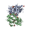
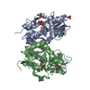
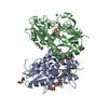
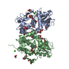
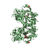
登録情報 データベース : PDB / ID : 3fatタイトル X-ray structure of iGluR4 flip ligand-binding core (S1S2) in complex with (S)-AMPA at 1.90A resolution Glutamate receptor 4 キーワード / / / / / 機能・相同性 分子機能 ドメイン・相同性 構成要素
/ / / / / / / / / / / / / / / / / / / / / / / / / / / / / / / / / / / / / / / / / / / / / / / / / / / / / / / / / / / / / / / 生物種 Rattus norvegicus (ドブネズミ)手法 / / / 解像度 : 1.9 Å データ登録者 Kasper, C. / Frydenvang, K. / Naur, P. / Gajhede, M. / Kastrup, J.S. ジャーナル : Febs Lett. / 年 : 2008タイトル : Molecular mechanism of agonist recognition by the ligand-binding core of the ionotropic glutamate receptor 4著者 : Kasper, C. / Frydenvang, K. / Naur, P. / Gajhede, M. / Pickering, D.S. / Kastrup, J.S. 履歴 登録 2008年11月18日 登録サイト / 処理サイト 改定 1.0 2008年12月9日 Provider / タイプ 改定 1.1 2011年7月13日 Group / Version format compliance改定 1.2 2017年8月9日 Group / Refinement description / Source and taxonomyカテゴリ / entity_src_gen / software / Item 改定 1.3 2023年11月1日 Group Data collection / Database references ... Data collection / Database references / Derived calculations / Refinement description カテゴリ chem_comp_atom / chem_comp_bond ... chem_comp_atom / chem_comp_bond / database_2 / pdbx_initial_refinement_model / struct_ref_seq_dif / struct_site Item _database_2.pdbx_DOI / _database_2.pdbx_database_accession ... _database_2.pdbx_DOI / _database_2.pdbx_database_accession / _struct_ref_seq_dif.details / _struct_site.pdbx_auth_asym_id / _struct_site.pdbx_auth_comp_id / _struct_site.pdbx_auth_seq_id
すべて表示 表示を減らす
 ムービー
ムービー コントローラー


 データを開く
 基本情報
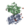
基本情報 要素
要素 グルタミン酸受容体
  キーワード
キーワード 機能・相同性情報
 データ登録者
 引用
引用 構造の表示
構造の表示 ダウンロードとリンク
 その他のダウンロード
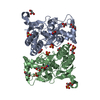


 PDBj
PDBj


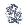
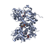
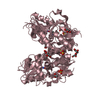
 集合体
集合体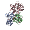

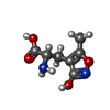


 分子量: 186.165 Da / 分子数: 3 / 由来タイプ: 合成 / 式: C7H10N2O4 / コメント: 神経伝達物質, アゴニスト*YM
 分子量: 96.063 Da / 分子数: 7 / 由来タイプ: 合成 / 式: SO4
 分子量: 92.094 Da / 分子数: 9 / 由来タイプ: 合成 / 式: C3H8O3
 分子量: 60.052 Da / 分子数: 6 / 由来タイプ: 合成 / 式: C2H4O2
 試料調製
試料調製 / ビームライン: I911-2 / 波長: 1.0412 Å
 解析
解析