- PDB-2xhd: Crystal structure of N-((2S)-5-(6-fluoro-3-pyridinyl)-2,3-dihydro... -
+
Open data
ID or keywords:
Loading...
-
Basic information
Entry
Database: PDB / ID: 2xhd
Title
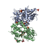
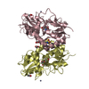
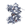
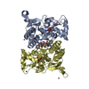
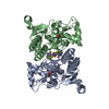
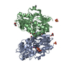
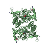
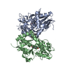
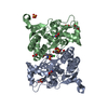
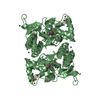
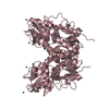
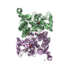
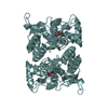
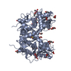
Crystal structure of N-((2S)-5-(6-fluoro-3-pyridinyl)-2,3-dihydro-1H- inden-2-yl)-2-propanesulfonamide in complex with the ligand binding domain of the human GluA2 receptor
Components
GLUTAMATE RECEPTOR 2GRIA2
Keywords
TRANSPORT PROTEIN / ION CHANNEL
Function / homology
Function and homology information
Activation of AMPA receptors / postsynaptic endocytic zone / Trafficking of GluR2-containing AMPA receptors / Unblocking of NMDA receptors, glutamate binding and activation / AMPA glutamate receptor activity / Long-term potentiation / AMPA glutamate receptor complex / excitatory synapse / asymmetric synapse / glutamate-gated receptor activity ...Activation of AMPA receptors / postsynaptic endocytic zone / Trafficking of GluR2-containing AMPA receptors / Unblocking of NMDA receptors, glutamate binding and activation / AMPA glutamate receptor activity / Long-term potentiation / AMPA glutamate receptor complex / excitatory synapse / asymmetric synapse / glutamate-gated receptor activity / MECP2 regulates neuronal receptors and channels / ionotropic glutamate receptor signaling pathway / transmitter-gated monoatomic ion channel activity involved in regulation of postsynaptic membrane potential / synaptic transmission, glutamatergic / postsynaptic density membrane / modulation of chemical synaptic transmission / endocytic vesicle membrane / amyloid-beta binding / chemical synaptic transmission / postsynapse / dendritic spine / postsynaptic density / external side of plasma membrane / neuronal cell body / dendrite / endoplasmic reticulum membrane / signal transduction / plasma membrane Similarity search - Function
Bacterial extracellular solute-binding proteins, family 3 / Solute-binding protein family 3/N-terminal domain of MltF / Ionotropic glutamate receptor, metazoa / Ligated ion channel L-glutamate- and glycine-binding site / : / Ligand-gated ion channel / Ionotropic glutamate receptor, L-glutamate and glycine-binding domain / Ligated ion channel L-glutamate- and glycine-binding site / Ionotropic glutamate receptor / Eukaryotic homologues of bacterial periplasmic substrate binding proteins. ...Bacterial extracellular solute-binding proteins, family 3 / Solute-binding protein family 3/N-terminal domain of MltF / Ionotropic glutamate receptor, metazoa / Ligated ion channel L-glutamate- and glycine-binding site / : / Ligand-gated ion channel / Ionotropic glutamate receptor, L-glutamate and glycine-binding domain / Ligated ion channel L-glutamate- and glycine-binding site / Ionotropic glutamate receptor / Eukaryotic homologues of bacterial periplasmic substrate binding proteins. / Receptor, ligand binding region / Receptor family ligand binding region / Periplasmic binding protein-like II / Periplasmic binding protein-like I / D-Maltodextrin-Binding Protein; domain 2 / 3-Layer(aba) Sandwich / Alpha Beta Similarity search - Domain/homology
Mass: 29277.814 Da / Num. of mol.: 2 Fragment: LIGAND BINDING DOMAIN, RESIDUES 412-427 AND 653-796 Source method: isolated from a genetically manipulated source Details: THIS IS AN S1-S2 FUSION IN WHICH GLY 118 AND THR 119 REPLACE A MEMBRANE SPANNING REGION. Source: (gene. exp.) HOMO SAPIENS (human) / Plasmid: PET15B / Production host: ESCHERICHIA COLI (E. coli) / Strain (production host): BL21(DE3) / References: UniProt: P42262
Mass: 18.015 Da / Num. of mol.: 545 / Source method: isolated from a natural source / Formula: H2O
Nonpolymer details
N-[(2S)-5-(6-FLUORO-3-PYRIDINYL)-2, 3-DIHYDRO-1H-INDEN-2-YL]-2-PROPANESULFONAMIDE (7T9): COMPOUND ...N-[(2S)-5-(6-FLUORO-3-PYRIDINYL)-2, 3-DIHYDRO-1H-INDEN-2-YL]-2-PROPANESULFONAMIDE (7T9): COMPOUND SITS ON NON-CRYSTALLOGRAPHIC TWOFOLD. TWO EQUIVALENT BINDING MODES ARE OBSEREVED. GLUTAMATE (GLU): GLUTAMATE IS THE LIGAND FOR THE RECEPTOR.
Sequence details
THIS IS AN S1-S2 FUSION IN WHICH GLY 118 AND THR 119 REPLACE A MEMBRANE SPANNING REGION.
-
Experimental details
-
Experiment
Experiment
Method: X-RAY DIFFRACTION / Number of used crystals: 1
-
Sample preparation
Crystal
Density Matthews: 2.45 Å3/Da / Density % sol: 49.75 % / Description: NONE
Protocol: SINGLE WAVELENGTH / Monochromatic (M) / Laue (L): M / Scattering type: x-ray
Radiation wavelength
Wavelength: 0.9393 Å / Relative weight: 1
Reflection
Resolution: 1.8→40 Å / Num. obs: 53509 / % possible obs: 99.4 % / Observed criterion σ(I): 0 / Redundancy: 4.3 % / Rmerge(I) obs: 0.07 / Net I/σ(I): 18.4
-
Processing
Software
Name
Version
Classification
REFMAC
5.5.0109
refinement
HKL
datareduction
SCALEPACK
datascaling
Refinement
Method to determine structure: OTHER Starting model: NONE Resolution: 1.8→20 Å / Cor.coef. Fo:Fc: 0.956 / Cor.coef. Fo:Fc free: 0.937 / SU B: 2.992 / SU ML: 0.093 / Cross valid method: THROUGHOUT / ESU R: 0.144 / ESU R Free: 0.135 / Stereochemistry target values: MAXIMUM LIKELIHOOD Details: HYDROGENS HAVE BEEN ADDED IN THE RIDING POSITIONS. COMPOUND 7T9 ON POCKET ON NCS TWOFOLD AXIS THE OCCUPANCY FOR THE COMPOUND AND SOME ASSOCIATED WATERS AND SIDE-CHAINS ARE 0.5. WATERS 3000- ...Details: HYDROGENS HAVE BEEN ADDED IN THE RIDING POSITIONS. COMPOUND 7T9 ON POCKET ON NCS TWOFOLD AXIS THE OCCUPANCY FOR THE COMPOUND AND SOME ASSOCIATED WATERS AND SIDE-CHAINS ARE 0.5. WATERS 3000-3003 ARE ASSOCIATED ASSOCIATED WITH CONFORMER B, 3004-3007 WITH CONFORMER A
Rfactor
Num. reflection
% reflection
Selection details
Rfree
0.238
2702
5.1 %
RANDOM
Rwork
0.2
-
-
-
obs
0.202
50730
100 %
-
Solvent computation
Ion probe radii: 0.8 Å / Shrinkage radii: 0.8 Å / VDW probe radii: 1.4 Å / Solvent model: BABINET MODEL WITH MASK
 Movie
Movie Controller

 Yorodumi
 Open data
 Basic information
 Components
 GRIA2
GRIA2  Keywords
 Function and homology information
 Authors
 Citation
 Structure visualization
 Downloads & links
 Other downloads


 PDBj
PDBj


 Assembly


 Type: L-peptide linking / Mass: 147.129 Da / Num. of mol.: 2 / Source method: obtained synthetically / Formula: C5H9NO4
 Mass: 96.063 Da / Num. of mol.: 5 / Source method: obtained synthetically / Formula: SO4
 Mass: 334.408 Da / Num. of mol.: 1 / Source method: obtained synthetically / Formula: C17H19FN2O2S
 Mass: 18.015 Da / Num. of mol.: 545 / Source method: isolated from a natural source / Formula: H2O
 Sample preparation
 / Beamline: ID23-1 / Wavelength: 0.9393
  Processing