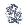
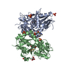
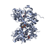
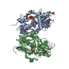
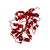
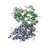
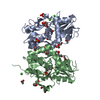
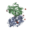
This entry contains the crystallographic asymmetric unit which consists of three chains. Chain A and B forms a dimer, whereas chain C forms a dimer with a symmetry related chain C (-x+1,y,-z+1). A complete tetrameric multimer representing the known biologically significant oligomerization state of the molecule cannot be generated by symmetry within the crystal.
Resolution: 1.9→32.644 Å / Occupancy max: 1 / Occupancy min: 0.5 / FOM work R set: 0.869 / SU ML: 0.26 / Cross valid method: THROUGHOUT / σ(F): 1.36 / Stereochemistry target values: Engh & Huber Details: Chain A: residue 260 was not observed in the electron density map Chain B: residues 256 and 260 were not observed in eletron density map
Rfactor
Num. reflection
% reflection
Selection details
Rfree
0.216
4371
5.01 %
random
Rwork
0.175
-
-
-
all
0.177
87195
-
-
obs
0.177
87195
99.85 %
-
Solvent computation
Shrinkage radii: 0.9 Å / VDW probe radii: 1.11 Å / Solvent model: FLAT BULK SOLVENT MODEL / Bsol: 41.02 Å2 / ksol: 0.355 e/Å3
In the structure databanks used in Yorodumi, some data are registered as the other names, "COVID-19 virus" and "2019-nCoV". Here are the details of the virus and the list of structure data.
Jan 31, 2019. EMDB accession codes are about to change! (news from PDBe EMDB page)
EMDB accession codes are about to change! (news from PDBe EMDB page)
The allocation of 4 digits for EMDB accession codes will soon come to an end. Whilst these codes will remain in use, new EMDB accession codes will include an additional digit and will expand incrementally as the available range of codes is exhausted. The current 4-digit format prefixed with “EMD-” (i.e. EMD-XXXX) will advance to a 5-digit format (i.e. EMD-XXXXX), and so on. It is currently estimated that the 4-digit codes will be depleted around Spring 2019, at which point the 5-digit format will come into force.
The EM Navigator/Yorodumi systems omit the EMD- prefix.
Related info.:Q: What is EMD? / ID/Accession-code notation in Yorodumi/EM Navigator
Yorodumi is a browser for structure data from EMDB, PDB, SASBDB, etc.
This page is also the successor to EM Navigator detail page, and also detail information page/front-end page for Omokage search.
The word "yorodu" (or yorozu) is an old Japanese word meaning "ten thousand". "mi" (miru) is to see.
Related info.:EMDB / PDB / SASBDB / Comparison of 3 databanks / Yorodumi Search / Aug 31, 2016. New EM Navigator & Yorodumi / Yorodumi Papers / Jmol/JSmol / Function and homology information / Changes in new EM Navigator and Yorodumi
 Movie
Movie Controller

 Yorodumi
 Open data
 Basic information
 Components
 Keywords
 Function and homology information
 Authors
 Citation
 Structure visualization
 Downloads & links
 Other downloads
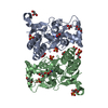


 PDBj
PDBj


 Assembly
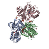


 Mass: 186.165 Da / Num. of mol.: 3 / Source method: obtained synthetically / Formula: C7H10N2O4 / Comment: neurotransmitter, agonist*YM
 Mass: 96.063 Da / Num. of mol.: 7 / Source method: obtained synthetically / Formula: SO4
 Mass: 92.094 Da / Num. of mol.: 9 / Source method: obtained synthetically / Formula: C3H8O3
 Mass: 60.052 Da / Num. of mol.: 6 / Source method: obtained synthetically / Formula: C2H4O2
 Sample preparation
 / Beamline: I911-2 / Wavelength: 1.0412 Å
 Processing