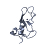
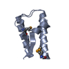
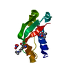
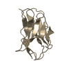
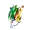
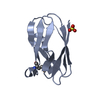
Journal: J.Biol.Inorg.Chem. / Year: 2009 Title: Insight into the protein and solvent contributions to the reduction potentials of [4Fe-4S]2+/+ clusters: crystal structures of the Allochromatium vinosum ferredoxin variants C57A and V13G and ...Title: Insight into the protein and solvent contributions to the reduction potentials of [4Fe-4S]2+/+ clusters: crystal structures of the Allochromatium vinosum ferredoxin variants C57A and V13G and the homologous Escherichia coli ferredoxin. Authors: Saridakis, E. / Giastas, P. / Efthymiou, G. / Thoma, V. / Moulis, J.M. / Kyritsis, P. / Mavridis, I.M.
Resolution: 1.05→14 Å / Num. parameters: 8225 / Num. restraintsaints: 9285 / Cross valid method: FREE R / σ(F): 0 / Stereochemistry target values: ENGH AND HUBER Details: ANISOTROPIC REFINEMENT REDUCED FREE R (NO CUTOFF). THE MISSING R-WORK VALUE IS DUE TO THE FACT THAT THE DATA OF THE REFERENCE AND WORKING SETS WERE MERGED IN THE LAST 6 CYCLES OF SHELX ...Details: ANISOTROPIC REFINEMENT REDUCED FREE R (NO CUTOFF). THE MISSING R-WORK VALUE IS DUE TO THE FACT THAT THE DATA OF THE REFERENCE AND WORKING SETS WERE MERGED IN THE LAST 6 CYCLES OF SHELX REFINEMENT, SO THERE IS ONLY ONE FINAL R (R = 0.1028 FOR FO>4SIG(FO) AND 0.1159 FOR ALL DATA).
Rfactor
Num. reflection
Selection details
all
0.116
54527
-
obs
0.103
41185
-
Rfree
-
-
RANDOM
Refine analyze
Num. disordered residues: 10 / Occupancy sum hydrogen: 0 / Occupancy sum non hydrogen: 812.97
Refinement step
Cycle: LAST / Resolution: 1.05→14 Å
Protein
Nucleic acid
Ligand
Solvent
Total
Num. atoms
651
0
16
246
913
Refine LS restraints
Refine-ID
Type
Dev ideal
X-RAY DIFFRACTION
s_bond_d
0.02
X-RAY DIFFRACTION
s_angle_d
0.044
X-RAY DIFFRACTION
s_similar_dist
X-RAY DIFFRACTION
s_from_restr_planes
X-RAY DIFFRACTION
s_zero_chiral_vol
X-RAY DIFFRACTION
s_non_zero_chiral_vol
X-RAY DIFFRACTION
s_anti_bump_dis_restr
0.034
X-RAY DIFFRACTION
s_rigid_bond_adp_cmpnt
X-RAY DIFFRACTION
s_similar_adp_cmpnt
X-RAY DIFFRACTION
s_approx_iso_adps
+
About Yorodumi
-
News
-
Feb 9, 2022. New format data for meta-information of EMDB entries
New format data for meta-information of EMDB entries
Version 3 of the EMDB header file is now the official format.
The previous official version 1.9 will be removed from the archive.
In the structure databanks used in Yorodumi, some data are registered as the other names, "COVID-19 virus" and "2019-nCoV". Here are the details of the virus and the list of structure data.
Jan 31, 2019. EMDB accession codes are about to change! (news from PDBe EMDB page)
EMDB accession codes are about to change! (news from PDBe EMDB page)
The allocation of 4 digits for EMDB accession codes will soon come to an end. Whilst these codes will remain in use, new EMDB accession codes will include an additional digit and will expand incrementally as the available range of codes is exhausted. The current 4-digit format prefixed with “EMD-” (i.e. EMD-XXXX) will advance to a 5-digit format (i.e. EMD-XXXXX), and so on. It is currently estimated that the 4-digit codes will be depleted around Spring 2019, at which point the 5-digit format will come into force.
The EM Navigator/Yorodumi systems omit the EMD- prefix.
Related info.:Q: What is EMD? / ID/Accession-code notation in Yorodumi/EM Navigator
Yorodumi is a browser for structure data from EMDB, PDB, SASBDB, etc.
This page is also the successor to EM Navigator detail page, and also detail information page/front-end page for Omokage search.
The word "yorodu" (or yorozu) is an old Japanese word meaning "ten thousand". "mi" (miru) is to see.
Related info.:EMDB / PDB / SASBDB / Comparison of 3 databanks / Yorodumi Search / Aug 31, 2016. New EM Navigator & Yorodumi / Yorodumi Papers / Jmol/JSmol / Function and homology information / Changes in new EM Navigator and Yorodumi
 Movie
Movie Controller

 Yorodumi
 Open data
 Basic information
 Components
 Keywords
 Function and homology information
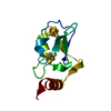
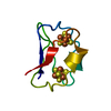
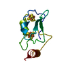
 Allochromatium vinosum (bacteria)
 Authors
 Citation
 Structure visualization
 Downloads & links
 Other downloads
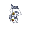


 PDBj
PDBj


 Assembly

 Mass: 351.640 Da / Num. of mol.: 2 / Source method: obtained synthetically / Formula: Fe4S4
 Mass: 18.015 Da / Num. of mol.: 245 / Source method: isolated from a natural source / Formula: H2O
 Sample preparation
 / Beamline: X13 / Wavelength: 0.8081
  Processing