 Movie
Movie Controller

[English] 日本語
 Yorodumi
- PDB-3eu5: Crystal structure of FTase(ALPHA-subunit; BETA-subunit DELTA C10)... -
+ Open data

- Basic information
| Entry | Database: PDB / ID: 3eu5 | ||||||
|---|---|---|---|---|---|---|---|
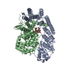
| Title | Crystal structure of FTase(ALPHA-subunit; BETA-subunit DELTA C10) in complex with BiotinGPP | ||||||
 Components Components |
| ||||||
 Keywords Keywords |  TRANSFERASE / protein prenylation / prenylome analysis / Prenyltransferase / Metal-binding / Phosphoprotein / Zinc TRANSFERASE / protein prenylation / prenylome analysis / Prenyltransferase / Metal-binding / Phosphoprotein / Zinc | ||||||
| Function / homology |  Function and homology information Function and homology informationApoptotic cleavage of cellular proteins / Inactivation, recovery and regulation of the phototransduction cascade / RAS processing / protein geranylgeranyltransferase activity / CAAX-protein geranylgeranyltransferase activity / CAAX-protein geranylgeranyltransferase complex / protein geranylgeranyltransferase type I / protein farnesylation / regulation of fibroblast proliferation / positive regulation of tubulin deacetylation ...Apoptotic cleavage of cellular proteins / Inactivation, recovery and regulation of the phototransduction cascade / RAS processing / protein geranylgeranyltransferase activity / CAAX-protein geranylgeranyltransferase activity / CAAX-protein geranylgeranyltransferase complex / protein geranylgeranyltransferase type I / protein farnesylation / regulation of fibroblast proliferation / positive regulation of tubulin deacetylation / protein farnesyltransferase / protein farnesyltransferase activity / protein farnesyltransferase complex / Rab geranylgeranyltransferase activity / protein geranylgeranylation / positive regulation of skeletal muscle acetylcholine-gated channel clustering / farnesyltranstransferase activity / negative regulation of nitric-oxide synthase biosynthetic process / microtubule associated complex / enzyme-linked receptor protein signaling pathway / positive regulation of nitric-oxide synthase biosynthetic process / positive regulation of Rac protein signal transduction / alpha-tubulin binding / response to inorganic substance / positive regulation of cell cycle / response to cytokine / wound healing / lipid metabolic process / response to organic cyclic compound / receptor tyrosine kinase binding / positive regulation of fibroblast proliferation / fibroblast proliferation / microtubule binding / cell population proliferation / molecular adaptor activity / negative regulation of cell population proliferation / positive regulation of cell population proliferation / negative regulation of apoptotic process / zinc ion binding / cytoplasmSimilarity search - Function | ||||||
| Biological species |  Rattus norvegicus (Norway rat) Rattus norvegicus (Norway rat) | ||||||
| Method | X-RAY DIFFRACTION / SYNCHROTRON / MOLECULAR REPLACEMENT / Resolution: 2.8 Å | ||||||
 Authors Authors | Guo, Z. / Nguyen, U.T.T. / Delon, C. / Bon, R.S. / Blankenfeldt, W. / Goody, R.S. / Waldmann, H. / Wolters, D. / Alexandrov, K. | ||||||
 Citation Citation | Journal: Nat.Chem.Biol. / Year: 2009 Title: Analysis of the eukaryotic prenylome by isoprenoid affinity tagging Authors: Nguyen, U.T.T. / Guo, Z. / Delon, C. / Wu, Y. / Deraeve, C. / Franzel, B. / Bon, R.S. / Blankenfeldt, W. / Goody, R.S. / Waldmann, H. / Wolters, D. / Alexandrov, K. | ||||||
| History |
|
- Structure visualization
| Structure viewer | Molecule: MolmilJmol/JSmol |
|---|
- Downloads & links
-Download
| PDBx/mmCIF format | 3eu5.cif.gz | 307.2 KB | Display | PDBx/mmCIF format |
|---|---|---|---|---|
| PDB format | pdb3eu5.ent.gz | 252.6 KB | Display | PDB format |
| PDBx/mmJSON format | 3eu5.json.gz | Tree view | PDBx/mmJSON format | |
| Others |  Other downloads Other downloads |
-Validation report
| Arichive directory | https://data.pdbj.org/pub/pdb/validation_reports/eu/3eu5ftp://data.pdbj.org/pub/pdb/validation_reports/eu/3eu5 | HTTPS FTP |
|---|
-Related structure data
| Related structure data |  3euvC  1ft1S C: citing same article ( S: Starting model for refinement |
|---|---|
| Similar structure data |


-Links
 PDBj
PDBj

- Assembly
| Deposited unit | 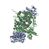
| ||||||||
|---|---|---|---|---|---|---|---|---|---|
| 1 |
| ||||||||
| Unit cell |
|
-Components
| #1: Protein | Mass: 44098.145 Da / Num. of mol.: 1 Source method: isolated from a genetically manipulated source Details: coexpression of alpha-subunit from pGATEV and engineered beta-subunit from pET27b Source: (gene. exp.) Rattus norvegicus (Norway rat) / Plasmid: pGATEV / Production host:  Escherichia coli (E. coli) / Strain (production host): BL21 CODON-PLUS RIL (DE3) Escherichia coli (E. coli) / Strain (production host): BL21 CODON-PLUS RIL (DE3)References: UniProt: Q04631, protein farnesyltransferase, protein geranylgeranyltransferase type I |
|---|---|
| #2: Protein | Farnesyltransferase / FTase-beta / CAAX farnesyltransferase subunit beta / RAS proteins prenyltransferase beta Mass: 47749.340 Da / Num. of mol.: 1 Fragment: DELTA C-terminus 10 AA, RABGGTase BETA-subunit, UNP residues 1-427 Source method: isolated from a genetically manipulated source Details: coexpression of alpha-subunit from pGATEV and engineered beta-subunit from pET27b Source: (gene. exp.) Rattus norvegicus (Norway rat) / Plasmid: pET27b / Production host: Escherichia coli (E. coli) / Strain (production host): BL21 CODON-PLUS RIL (DE3) / References: UniProt: Q02293, protein farnesyltransferase |
| #3: Chemical | ChemComp-ZN /   Mass: 65.409 Da / Num. of mol.: 1 / Source method: obtained synthetically / Formula: Zn Mass: 65.409 Da / Num. of mol.: 1 / Source method: obtained synthetically / Formula: Zn |
| #4: Chemical | ChemComp-GBO / (  Mass: 555.519 Da / Num. of mol.: 1 / Source method: obtained synthetically / Formula: C20H35N3O9P2S Mass: 555.519 Da / Num. of mol.: 1 / Source method: obtained synthetically / Formula: C20H35N3O9P2S |
| #5: Water | ChemComp-HOH / Water Mass: 18.015 Da / Num. of mol.: 132 / Source method: isolated from a natural source / Formula: H2O Mass: 18.015 Da / Num. of mol.: 132 / Source method: isolated from a natural source / Formula: H2O |
-Experimental details
-Experiment
| Experiment | Method: X-RAY DIFFRACTION / Number of used crystals: 1 |
|---|
- Sample preparation
| Crystal | Density Matthews: 3.4 Å3/Da / Density % sol: 63.84 % |
|---|---|
| Crystal grow | Temperature: 292 K / Method: vapor diffusion, hanging drop / pH: 7.5 Details: 20% (w/v) PEG 4000, 0.2M MgCl2, 0.1M Tris, pH 7.5, VAPOR DIFFUSION, HANGING DROP, temperature 292K |
-Data collection
| Diffraction | Mean temperature: 100 K |
|---|---|
| Diffraction source | Source: SYNCHROTRON / Site: SLS  / Beamline: X10SA / Wavelength: 1.0007 Å / Beamline: X10SA / Wavelength: 1.0007 Å |
| Detector | Type: MARMOSAIC 225 mm CCD / Detector: CCD / Date: May 25, 2008 |
| Radiation | Monochromator: SI(111) / Protocol: SINGLE WAVELENGTH / Monochromatic (M) / Laue (L): M / Scattering type: x-ray |
| Radiation wavelength | Wavelength: 1.0007 Å / Relative weight: 1 |
| Reflection | Resolution: 2.8→30 Å / Num. all: 30693 / Num. obs: 30615 / % possible obs: 99.7 % / Observed criterion σ(I): 5 / Redundancy: 7.6 % / Biso Wilson estimate: 53 Å2 / Rsym value: 0.069 / Net I/σ(I): 20.5 |
| Reflection shell | Resolution: 2.8→2.9 Å / Redundancy: 7.6 % / Mean I/σ(I) obs: 5.9 / Num. unique all: 30615 / Rsym value: 0.335 / % possible all: 99.8 |
- Processing
| Software |
| ||||||||||||||||||||||||||||||||||||||||||||||||||||||||||||||||||||||||||||||||||||||||||||||||||||||||||||||||||||||||||||||||||||||||||||||||||||||
|---|---|---|---|---|---|---|---|---|---|---|---|---|---|---|---|---|---|---|---|---|---|---|---|---|---|---|---|---|---|---|---|---|---|---|---|---|---|---|---|---|---|---|---|---|---|---|---|---|---|---|---|---|---|---|---|---|---|---|---|---|---|---|---|---|---|---|---|---|---|---|---|---|---|---|---|---|---|---|---|---|---|---|---|---|---|---|---|---|---|---|---|---|---|---|---|---|---|---|---|---|---|---|---|---|---|---|---|---|---|---|---|---|---|---|---|---|---|---|---|---|---|---|---|---|---|---|---|---|---|---|---|---|---|---|---|---|---|---|---|---|---|---|---|---|---|---|---|---|---|---|---|
| Refinement | Method to determine structure: MOLECULAR REPLACEMENT Starting model: PDB ENTRY 1FT1 Resolution: 2.8→29.96 Å / Cor.coef. Fo:Fc: 0.963 / Cor.coef. Fo:Fc free: 0.931 / SU B: 21.465 / SU ML: 0.186 / Cross valid method: THROUGHOUT / ESU R Free: 0.269 / Stereochemistry target values: MAXIMUM LIKELIHOOD / Details: HYDROGENS HAVE BEEN ADDED IN THE RIDING POSITIONS
| ||||||||||||||||||||||||||||||||||||||||||||||||||||||||||||||||||||||||||||||||||||||||||||||||||||||||||||||||||||||||||||||||||||||||||||||||||||||
| Solvent computation | Ion probe radii: 0.8 Å / Shrinkage radii: 0.8 Å / VDW probe radii: 1.2 Å / Solvent model: BABINET MODEL WITH MASK | ||||||||||||||||||||||||||||||||||||||||||||||||||||||||||||||||||||||||||||||||||||||||||||||||||||||||||||||||||||||||||||||||||||||||||||||||||||||
| Displacement parameters | Biso mean: 52.562 Å2
| ||||||||||||||||||||||||||||||||||||||||||||||||||||||||||||||||||||||||||||||||||||||||||||||||||||||||||||||||||||||||||||||||||||||||||||||||||||||
| Refinement step | Cycle: LAST / Resolution: 2.8→29.96 Å
| ||||||||||||||||||||||||||||||||||||||||||||||||||||||||||||||||||||||||||||||||||||||||||||||||||||||||||||||||||||||||||||||||||||||||||||||||||||||
| Refine LS restraints |
| ||||||||||||||||||||||||||||||||||||||||||||||||||||||||||||||||||||||||||||||||||||||||||||||||||||||||||||||||||||||||||||||||||||||||||||||||||||||
| LS refinement shell | Resolution: 2.8→2.872 Å / Total num. of bins used: 20
| ||||||||||||||||||||||||||||||||||||||||||||||||||||||||||||||||||||||||||||||||||||||||||||||||||||||||||||||||||||||||||||||||||||||||||||||||||||||
| Refinement TLS params. | Method: refined / Refine-ID: X-RAY DIFFRACTION
| ||||||||||||||||||||||||||||||||||||||||||||||||||||||||||||||||||||||||||||||||||||||||||||||||||||||||||||||||||||||||||||||||||||||||||||||||||||||
| Refinement TLS group |
|
