 Movie
Movie Controller

[English] 日本語
 Yorodumi
- PDB-3ek9: SPRY Domain-containing SOCS Box Protein 2: Crystal Structure and ... -
+ Open data

- Basic information
| Entry | Database: PDB / ID: 3ek9 | ||||||
|---|---|---|---|---|---|---|---|
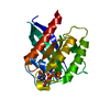
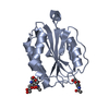
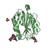
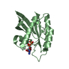
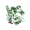
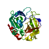
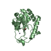
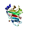
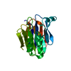
| Title | SPRY Domain-containing SOCS Box Protein 2: Crystal Structure and Residues Critical for Protein Binding | ||||||
 Components Components | SPRY domain-containing SOCS box protein 2 | ||||||
 Keywords Keywords |  PROTEIN BINDING / SPRY domain / Ubl conjugation pathway / SIGNALING PROTEIN PROTEIN BINDING / SPRY domain / Ubl conjugation pathway / SIGNALING PROTEIN | ||||||
| Function / homology |  Function and homology informationNeddylation / Antigen processing: Ubiquitination & Proteasome degradation / SCF ubiquitin ligase complex / ubiquitin-like ligase-substrate adaptor activity / ubiquitin-dependent protein catabolic process / proteasome-mediated ubiquitin-dependent protein catabolic process / protein ubiquitination / intracellular signal transduction / cytosol Function and homology informationNeddylation / Antigen processing: Ubiquitination & Proteasome degradation / SCF ubiquitin ligase complex / ubiquitin-like ligase-substrate adaptor activity / ubiquitin-dependent protein catabolic process / proteasome-mediated ubiquitin-dependent protein catabolic process / protein ubiquitination / intracellular signal transduction / cytosolSimilarity search - Function | ||||||
| Biological species |  Mus musculus (house mouse) Mus musculus (house mouse) | ||||||
| Method | X-RAY DIFFRACTION / MOLECULAR REPLACEMENT / Resolution: 2.6 Å | ||||||
 Authors Authors | Kuang, Z. / Yao, S. / Xu, Y. / Garrett, T.J.P. / Norton, R.S. | ||||||
 Citation Citation | Journal: J.Mol.Biol. / Year: 2009 Title: SPRY domain-containing SOCS box protein 2: crystal structure and residues critical for protein binding. Authors: Kuang, Z. / Yao, S. / Xu, Y. / Lewis, R.S. / Low, A. / Masters, S.L. / Willson, T.A. / Kolesnik, T.B. / Nicholson, S.E. / Garrett, T.J. / Norton, R.S. | ||||||
| History |
|
- Structure visualization
| Structure viewer | Molecule: MolmilJmol/JSmol |
|---|
- Downloads & links
-Download
| PDBx/mmCIF format | 3ek9.cif.gz | 52.4 KB | Display | PDBx/mmCIF format |
|---|---|---|---|---|
| PDB format | pdb3ek9.ent.gz | 35.8 KB | Display | PDB format |
| PDBx/mmJSON format | 3ek9.json.gz | Tree view | PDBx/mmJSON format | |
| Others |  Other downloads Other downloads |
-Validation report
| Arichive directory | https://data.pdbj.org/pub/pdb/validation_reports/ek/3ek9ftp://data.pdbj.org/pub/pdb/validation_reports/ek/3ek9 | HTTPS FTP |
|---|
-Related structure data
| Related structure data | 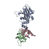 2fnjS S: Starting model for refinement |
|---|---|
| Similar structure data |

-Links
 PDBj
PDBj


- Assembly
| Deposited unit | 
| ||||||||
|---|---|---|---|---|---|---|---|---|---|
| 1 |
| ||||||||
| Unit cell |
|
-Components
| #1: Protein | Mass: 23365.156 Da / Num. of mol.: 1 / Fragment: B30.2/SPRY domain residues 12-224 Source method: isolated from a genetically manipulated source Source: (gene. exp.) Mus musculus (house mouse) / Gene: Spsb2, Grcc9, Ssb2 / Production host:  Escherichia coli (E. coli) / References: UniProt: O88838 Escherichia coli (E. coli) / References: UniProt: O88838 |
|---|---|
| #2: Chemical | ChemComp-GOL / Glycerol  Mass: 92.094 Da / Num. of mol.: 1 / Source method: obtained synthetically / Formula: C3H8O3 Mass: 92.094 Da / Num. of mol.: 1 / Source method: obtained synthetically / Formula: C3H8O3 |
| #3: Water | ChemComp-HOH / Water Mass: 18.015 Da / Num. of mol.: 59 / Source method: isolated from a natural source / Formula: H2O Mass: 18.015 Da / Num. of mol.: 59 / Source method: isolated from a natural source / Formula: H2O |
-Experimental details
-Experiment
| Experiment | Method: X-RAY DIFFRACTION / Number of used crystals: 1 |
|---|
- Sample preparation
| Crystal grow | Temperature: 293 K / Method: evaporation / pH: 6.5 Details: 100mM Bis-Tris, pH6.5, 200mM MgCl2, 25% PEG3350, EVAPORATION, temperature 293K |
|---|
-Data collection
| Diffraction | Mean temperature: 100 K |
|---|---|
| Diffraction source | Source: ROTATING ANODE / Type: RIGAKU MICROMAX-007 / Wavelength: 1.54 Å |
| Detector | Type: RIGAKU RAXIS IV / Detector: IMAGE PLATE / Date: Jun 13, 2007 / Details: AXCO |
| Radiation | Protocol: SINGLE WAVELENGTH / Monochromatic (M) / Laue (L): M / Scattering type: x-ray |
| Radiation wavelength | Wavelength: 1.54 Å / Relative weight: 1 |
| Reflection | Resolution: 2.6→30.7 Å / Num. all: 5018 / Num. obs: 5002 / % possible obs: 94 % / Observed criterion σ(F): 2 / Observed criterion σ(I): 1 / Redundancy: 3.7 % / Rmerge(I) obs: 0.117 |
| Reflection shell | Resolution: 2.6→2.69 Å / Redundancy: 3.3 % / Rmerge(I) obs: 0.356 / Mean I/σ(I) obs: 3.2 / Num. unique all: 493 / % possible all: 93.9 |
- Processing
| Software |
| |||||||||||||||||||||||||||||||||||||||||||||||||||||||||||||||||
|---|---|---|---|---|---|---|---|---|---|---|---|---|---|---|---|---|---|---|---|---|---|---|---|---|---|---|---|---|---|---|---|---|---|---|---|---|---|---|---|---|---|---|---|---|---|---|---|---|---|---|---|---|---|---|---|---|---|---|---|---|---|---|---|---|---|---|
| Refinement | Method to determine structure: MOLECULAR REPLACEMENT Starting model: PDB entry 2FNJ Resolution: 2.6→30.69 Å / Cor.coef. Fo:Fc: 0.945 / Cor.coef. Fo:Fc free: 0.833 / SU B: 12.125 / SU ML: 0.262 / Cross valid method: THROUGHOUT / ESU R Free: 0.419 / Stereochemistry target values: MAXIMUM LIKELIHOOD
| |||||||||||||||||||||||||||||||||||||||||||||||||||||||||||||||||
| Solvent computation | Ion probe radii: 0.8 Å / Shrinkage radii: 0.8 Å / VDW probe radii: 1.4 Å / Solvent model: MASK | |||||||||||||||||||||||||||||||||||||||||||||||||||||||||||||||||
| Displacement parameters | Biso mean: 17.309 Å2
| |||||||||||||||||||||||||||||||||||||||||||||||||||||||||||||||||
| Refinement step | Cycle: LAST / Resolution: 2.6→30.69 Å
| |||||||||||||||||||||||||||||||||||||||||||||||||||||||||||||||||
| Refine LS restraints |
| |||||||||||||||||||||||||||||||||||||||||||||||||||||||||||||||||
| LS refinement shell | Resolution: 2.6→2.67 Å / Total num. of bins used: 20
|
