 Movie
Movie Controller

[English] 日本語
 Yorodumi
- PDB-3ef5: Structure of the RNA pyrophosphohydrolase BdRppH in complex with dGTP -
+ Open data

- Basic information
| Entry | Database: PDB / ID: 3ef5 | ||||||
|---|---|---|---|---|---|---|---|
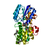
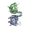
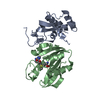
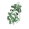
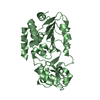
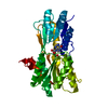
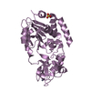
| Title | Structure of the RNA pyrophosphohydrolase BdRppH in complex with dGTP | ||||||
 Components Components | Probable pyrophosphohydrolase | ||||||
 Keywords Keywords |  HYDROLASE / Nudix / RNA pyrophosphohydrolase HYDROLASE / Nudix / RNA pyrophosphohydrolase | ||||||
| Function / homology |  Function and homology information8-oxo-dGTP diphosphatase / 8-oxo-7,8-dihydrodeoxyguanosine triphosphate pyrophosphatase activity / 8-oxo-7,8-dihydroguanosine triphosphate pyrophosphatase activity / DNA replication / DNA repair / GTP binding / identical protein binding / metal ion binding Function and homology information8-oxo-dGTP diphosphatase / 8-oxo-7,8-dihydrodeoxyguanosine triphosphate pyrophosphatase activity / 8-oxo-7,8-dihydroguanosine triphosphate pyrophosphatase activity / DNA replication / DNA repair / GTP binding / identical protein binding / metal ion bindingSimilarity search - Function | ||||||
| Biological species |  Bdellovibrio bacteriovorus (bacteria) Bdellovibrio bacteriovorus (bacteria) | ||||||
| Method | X-RAY DIFFRACTION / SYNCHROTRON / Resolution: 2.6 Å | ||||||
 Authors Authors | Messing, S.A. / Gabelli, S.B. / Amzel, L.M. | ||||||
 Citation Citation | Journal: Structure / Year: 2009 Title: Structure and Biological Function of the RNA Pyrophosphohydrolase BdRppH from Bdellovibrio bacteriovorus. Authors: Messing, S.A. / Gabelli, S.B. / Liu, Q. / Celesnik, H. / Belasco, J.G. / Pineiro, S.A. / Amzel, L.M. | ||||||
| History |
|
- Structure visualization
| Structure viewer | Molecule: MolmilJmol/JSmol |
|---|
- Downloads & links
-Download
| PDBx/mmCIF format | 3ef5.cif.gz | 68 KB | Display | PDBx/mmCIF format |
|---|---|---|---|---|
| PDB format | pdb3ef5.ent.gz | 49.9 KB | Display | PDB format |
| PDBx/mmJSON format | 3ef5.json.gz | Tree view | PDBx/mmJSON format | |
| Others |  Other downloads Other downloads |
-Validation report
| Arichive directory | https://data.pdbj.org/pub/pdb/validation_reports/ef/3ef5ftp://data.pdbj.org/pub/pdb/validation_reports/ef/3ef5 | HTTPS FTP |
|---|
-Related structure data


-Links
 PDBj
PDBj

- Assembly
| Deposited unit | 
| ||||||||
|---|---|---|---|---|---|---|---|---|---|
| 1 |
| ||||||||
| Unit cell |
| ||||||||
| Details | Gel filtration was used to determine the biological assembly. |
-Components
| #1: Protein | Mass: 17634.312 Da / Num. of mol.: 2 Source method: isolated from a genetically manipulated source Source: (gene. exp.) Bdellovibrio bacteriovorus (bacteria) / Strain: HD100 / Gene: mutT, Bd0714 / Plasmid: pET24a / Production host: Escherichia coli (E. coli) / Strain (production host): BL21(DE3)References: UniProt: Q6MPX4, Hydrolases; Acting on acid anhydrides; In phosphorus-containing anhydrides#2: Chemical | Deoxyguanosine triphosphate  Mass: 507.181 Da / Num. of mol.: 2 / Source method: obtained synthetically / Formula: C10H16N5O13P3 Mass: 507.181 Da / Num. of mol.: 2 / Source method: obtained synthetically / Formula: C10H16N5O13P3#3: Water | ChemComp-HOH / | Water Mass: 18.015 Da / Num. of mol.: 44 / Source method: isolated from a natural source / Formula: H2O Mass: 18.015 Da / Num. of mol.: 44 / Source method: isolated from a natural source / Formula: H2O |
|---|
-Experimental details
-Experiment
| Experiment | Method: X-RAY DIFFRACTION / Number of used crystals: 1 |
|---|
- Sample preparation
| Crystal | Density Matthews: 3.08 Å3/Da / Density % sol: 60.06 % |
|---|---|
| Crystal grow | Temperature: 298 K / Method: hanging drop / pH: 7 Details: PEG 4000, Na acetate, pH 7, hanging drop, temperature 298K |
-Data collection
| Diffraction | Mean temperature: 100 K | |||||||||||||||||||||||||||||||||||||||||||||||||||||||||||||||||||||||||||||
|---|---|---|---|---|---|---|---|---|---|---|---|---|---|---|---|---|---|---|---|---|---|---|---|---|---|---|---|---|---|---|---|---|---|---|---|---|---|---|---|---|---|---|---|---|---|---|---|---|---|---|---|---|---|---|---|---|---|---|---|---|---|---|---|---|---|---|---|---|---|---|---|---|---|---|---|---|---|---|
| Diffraction source | Source: SYNCHROTRON / Site: NSLS  / Beamline: X6A / Wavelength: 1 Å / Beamline: X6A / Wavelength: 1 Å | |||||||||||||||||||||||||||||||||||||||||||||||||||||||||||||||||||||||||||||
| Detector | Type: adsc Q210 / Detector: CCD / Date: Aug 16, 2008 | |||||||||||||||||||||||||||||||||||||||||||||||||||||||||||||||||||||||||||||
| Radiation | Protocol: SINGLE WAVELENGTH / Monochromatic (M) / Laue (L): M / Scattering type: x-ray | |||||||||||||||||||||||||||||||||||||||||||||||||||||||||||||||||||||||||||||
| Radiation wavelength | Wavelength: 1 Å / Relative weight: 1 | |||||||||||||||||||||||||||||||||||||||||||||||||||||||||||||||||||||||||||||
| Reflection | Resolution: 2.4→46.37 Å / Num. obs: 15730 / % possible obs: 89.2 % / Observed criterion σ(F): 0 / Observed criterion σ(I): 0 / Redundancy: 6.6 % / Rmerge(I) obs: 0.049 / Χ2: 0.827 / Net I/σ(I): 32.267 | |||||||||||||||||||||||||||||||||||||||||||||||||||||||||||||||||||||||||||||
| Reflection shell |
|
- Processing
| Software |
| |||||||||||||||||||||||||||||||||||||||||||||||||||||||||||||||||||||||||||||||||||||||||||||||||||||||||||||||||||||||||||||
|---|---|---|---|---|---|---|---|---|---|---|---|---|---|---|---|---|---|---|---|---|---|---|---|---|---|---|---|---|---|---|---|---|---|---|---|---|---|---|---|---|---|---|---|---|---|---|---|---|---|---|---|---|---|---|---|---|---|---|---|---|---|---|---|---|---|---|---|---|---|---|---|---|---|---|---|---|---|---|---|---|---|---|---|---|---|---|---|---|---|---|---|---|---|---|---|---|---|---|---|---|---|---|---|---|---|---|---|---|---|---|---|---|---|---|---|---|---|---|---|---|---|---|---|---|---|---|
| Refinement | Resolution: 2.6→46.37 Å / Cor.coef. Fo:Fc: 0.929 / Cor.coef. Fo:Fc free: 0.881 / WRfactor Rfree: 0.3 / WRfactor Rwork: 0.244 / Occupancy max: 1 / Occupancy min: 0.5 / FOM work R set: 0.766 / SU B: 23.858 / SU ML: 0.255 / SU R Cruickshank DPI: 0.502 / SU Rfree: 0.344 / TLS residual ADP flag: LIKELY RESIDUAL / Cross valid method: THROUGHOUT / σ(F): 0 / ESU R: 0.473 / ESU R Free: 0.328 / Stereochemistry target values: MAXIMUM LIKELIHOOD / Details: HYDROGENS HAVE BEEN ADDED IN THE RIDING POSITIONS
| |||||||||||||||||||||||||||||||||||||||||||||||||||||||||||||||||||||||||||||||||||||||||||||||||||||||||||||||||||||||||||||
| Solvent computation | Ion probe radii: 0.8 Å / Shrinkage radii: 0.8 Å / VDW probe radii: 1.4 Å / Solvent model: MASK | |||||||||||||||||||||||||||||||||||||||||||||||||||||||||||||||||||||||||||||||||||||||||||||||||||||||||||||||||||||||||||||
| Displacement parameters | Biso max: 89.09 Å2 / Biso mean: 58.584 Å2 / Biso min: 46.61 Å2
| |||||||||||||||||||||||||||||||||||||||||||||||||||||||||||||||||||||||||||||||||||||||||||||||||||||||||||||||||||||||||||||
| Refinement step | Cycle: LAST / Resolution: 2.6→46.37 Å
| |||||||||||||||||||||||||||||||||||||||||||||||||||||||||||||||||||||||||||||||||||||||||||||||||||||||||||||||||||||||||||||
| Refine LS restraints |
| |||||||||||||||||||||||||||||||||||||||||||||||||||||||||||||||||||||||||||||||||||||||||||||||||||||||||||||||||||||||||||||
| LS refinement shell | Resolution: 2.6→2.667 Å / Total num. of bins used: 20
| |||||||||||||||||||||||||||||||||||||||||||||||||||||||||||||||||||||||||||||||||||||||||||||||||||||||||||||||||||||||||||||
| Refinement TLS params. | Method: refined / Refine-ID: X-RAY DIFFRACTION
| |||||||||||||||||||||||||||||||||||||||||||||||||||||||||||||||||||||||||||||||||||||||||||||||||||||||||||||||||||||||||||||
| Refinement TLS group |
|
