 Movie
Movie Controller

[English] 日本語
 Yorodumi
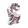
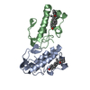
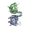
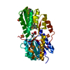
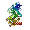
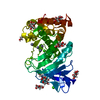
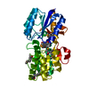
- PDB-3q87: Structure of E. cuniculi Mtq2-Trm112 complex responible for the m... -
+ Open data

- Basic information
| Entry | Database: PDB / ID: 3q87 | ||||||
|---|---|---|---|---|---|---|---|
| Title | Structure of E. cuniculi Mtq2-Trm112 complex responible for the methylation of eRF1 translation termination factor | ||||||
 Components Components |
| ||||||
 Keywords Keywords | TRANSFERASE ACTIVATOR/TRANSFERASE / SAM-methyltransferase /  methyltransferase / methylation / TRANSFERASE ACTIVATOR-TRANSFERASE complex methyltransferase / methylation / TRANSFERASE ACTIVATOR-TRANSFERASE complex | ||||||
| Function / homology |  Function and homology informationmethyltransferase activity / methylation / nucleic acid binding / metal ion binding Function and homology informationmethyltransferase activity / methylation / nucleic acid binding / metal ion bindingSimilarity search - Function | ||||||
| Biological species |  Encephalitozoon cuniculi (fungus) Encephalitozoon cuniculi (fungus) | ||||||
| Method | X-RAY DIFFRACTION / SYNCHROTRON / SAD / Resolution: 1.997 Å | ||||||
 Authors Authors | Liger, D. / Mora, L. / Lazar, N. / Figaro, S. / Henri, J. / Scrima, N. / Buckingham, R.H. / van Tilbeurgh, H. / Heurgue-Hamard, V. / Graille, M. | ||||||
 Citation Citation | Journal: Nucleic Acids Res. / Year: 2011 Title: Mechanism of activation of methyltransferases involved in translation by the Trm112 'hub' protein Authors: Liger, D. / Mora, L. / Lazar, N. / Figaro, S. / Henri, J. / Scrima, N. / Buckingham, R.H. / van Tilbeurgh, H. / Heurgue-Hamard, V. / Graille, M. | ||||||
| History |
|
- Structure visualization
| Structure viewer | Molecule: MolmilJmol/JSmol |
|---|
- Downloads & links
-Download
| PDBx/mmCIF format | 3q87.cif.gz | 73.7 KB | Display | PDBx/mmCIF format |
|---|---|---|---|---|
| PDB format | pdb3q87.ent.gz | 53.8 KB | Display | PDB format |
| PDBx/mmJSON format | 3q87.json.gz | Tree view | PDBx/mmJSON format | |
| Others |  Other downloads Other downloads |
-Validation report
| Arichive directory | https://data.pdbj.org/pub/pdb/validation_reports/q8/3q87ftp://data.pdbj.org/pub/pdb/validation_reports/q8/3q87 | HTTPS FTP |
|---|
-Related structure data
| Related structure data | |
|---|---|
| Similar structure data |


-Links
 PDBj
PDBj
- Assembly
| Deposited unit | 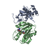
| ||||||||
|---|---|---|---|---|---|---|---|---|---|
| 1 |
| ||||||||
| Unit cell |
|
-Components
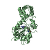
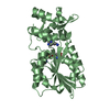
| #1: Protein | Mass: 14175.262 Da / Num. of mol.: 1 Source method: isolated from a genetically manipulated source Source: (gene. exp.) Encephalitozoon cuniculi (fungus) / Plasmid: pET21-a / Production host:  Escherichia coli (E. coli) / References: UniProt: Q8SUP0 Escherichia coli (E. coli) / References: UniProt: Q8SUP0 |
|---|---|
| #2: Protein | Mass: 19233.957 Da / Num. of mol.: 1 Source method: isolated from a genetically manipulated source Source: (gene. exp.) Encephalitozoon cuniculi (fungus) / Plasmid: pET21-a / Production host: Escherichia coli (E. coli) / References: UniProt: Q8SRR4 |
| #3: Chemical | ChemComp-ZN /   Mass: 65.409 Da / Num. of mol.: 1 / Source method: obtained synthetically / Formula: Zn Mass: 65.409 Da / Num. of mol.: 1 / Source method: obtained synthetically / Formula: Zn |
| #4: Chemical | ChemComp-SAM / S-Adenosyl methionine  Mass: 398.437 Da / Num. of mol.: 1 / Source method: obtained synthetically / Formula: C15H22N6O5S Mass: 398.437 Da / Num. of mol.: 1 / Source method: obtained synthetically / Formula: C15H22N6O5S |
| #5: Water | ChemComp-HOH / Water Mass: 18.015 Da / Num. of mol.: 136 / Source method: isolated from a natural source / Formula: H2O Mass: 18.015 Da / Num. of mol.: 136 / Source method: isolated from a natural source / Formula: H2O |
-Experimental details
-Experiment
| Experiment | Method: X-RAY DIFFRACTION / Number of used crystals: 1 |
|---|
- Sample preparation
| Crystal | Density Matthews: 2.27 Å3/Da / Density % sol: 45.72 % |
|---|---|
| Crystal grow | Temperature: 310 K / Method: vapor diffusion / pH: 7.5 Details: 20% PEG4000, 10% isopropanol, 100mM Hepes, pH 7.5, VAPOR DIFFUSION, temperature 310K |
-Data collection
| Diffraction | Mean temperature: 100 K |
|---|---|
| Diffraction source | Source: SYNCHROTRON / Site: SOLEIL  / Beamline: PROXIMA 1 / Wavelength: 1.214 Å / Beamline: PROXIMA 1 / Wavelength: 1.214 Å |
| Detector | Type: ADSC QUANTUM 315r / Detector: CCD / Date: Apr 20, 2008 |
| Radiation | Monochromator: Si 111 CHANNEL / Protocol: SINGLE WAVELENGTH / Monochromatic (M) / Laue (L): M / Scattering type: x-ray |
| Radiation wavelength | Wavelength: 1.214 Å / Relative weight: 1 |
| Reflection | Resolution: 1.997→50 Å / Num. all: 34474 / Num. obs: 33750 / % possible obs: 97.9 % / Observed criterion σ(F): 2 / Observed criterion σ(I): 2 / Redundancy: 2.4 % / Biso Wilson estimate: 24.64 Å2 / Rsym value: 0.089 / Net I/σ(I): 8.3 |
| Reflection shell | Resolution: 2→2.22 Å / Redundancy: 2.4 % / Mean I/σ(I) obs: 2 / Rsym value: 0.482 / % possible all: 96.9 |
- Processing
| Software |
| |||||||||||||||||||||||||||||||||||||||||||||||||||||||||||||||||||||||||||||||||||||||||||||||||||||||||
|---|---|---|---|---|---|---|---|---|---|---|---|---|---|---|---|---|---|---|---|---|---|---|---|---|---|---|---|---|---|---|---|---|---|---|---|---|---|---|---|---|---|---|---|---|---|---|---|---|---|---|---|---|---|---|---|---|---|---|---|---|---|---|---|---|---|---|---|---|---|---|---|---|---|---|---|---|---|---|---|---|---|---|---|---|---|---|---|---|---|---|---|---|---|---|---|---|---|---|---|---|---|---|---|---|---|---|
| Refinement | Method to determine structure: SAD / Resolution: 1.997→48.377 Å / Occupancy max: 1 / Occupancy min: 0.22 / FOM work R set: 0.8156 / SU ML: 0.31 / Cross valid method: THROUGHOUT / σ(F): 1.35 / Phase error: 25.68 / Stereochemistry target values: ML
| |||||||||||||||||||||||||||||||||||||||||||||||||||||||||||||||||||||||||||||||||||||||||||||||||||||||||
| Solvent computation | Shrinkage radii: 0.9 Å / VDW probe radii: 1.11 Å / Solvent model: FLAT BULK SOLVENT MODEL / Bsol: 42.901 Å2 / ksol: 0.349 e/Å3 | |||||||||||||||||||||||||||||||||||||||||||||||||||||||||||||||||||||||||||||||||||||||||||||||||||||||||
| Displacement parameters | Biso max: 102.19 Å2 / Biso mean: 30.2186 Å2 / Biso min: 9.96 Å2
| |||||||||||||||||||||||||||||||||||||||||||||||||||||||||||||||||||||||||||||||||||||||||||||||||||||||||
| Refinement step | Cycle: LAST / Resolution: 1.997→48.377 Å
| |||||||||||||||||||||||||||||||||||||||||||||||||||||||||||||||||||||||||||||||||||||||||||||||||||||||||
| Refine LS restraints |
| |||||||||||||||||||||||||||||||||||||||||||||||||||||||||||||||||||||||||||||||||||||||||||||||||||||||||
| LS refinement shell |
|
