 Movie
Movie Controller

[English] 日本語
 Yorodumi
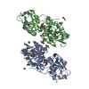
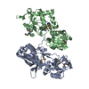
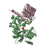
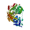
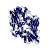
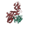
- PDB-3dsh: Crystal structure of dimeric interferon regulatory factor 5 (IRF-... -
+ Open data

- Basic information
| Entry | Database: PDB / ID: 3dsh | ||||||
|---|---|---|---|---|---|---|---|
| Title | Crystal structure of dimeric interferon regulatory factor 5 (IRF-5) transactivation domain | ||||||
 Components Components | Interferon regulatory factor 5 Interferon regulatory factors Interferon regulatory factors | ||||||
 Keywords Keywords | DNA BINDING PROTEIN / Phosphoactivation induced dimerization / DNA-binding / Nucleus / Transcription / Transcription regulation | ||||||
| Function / homology |  Function and homology information Function and homology informationresponse to peptidoglycan / chromatin => GO:0000785 / immune system process / type I interferon-mediated signaling pathway / positive regulation of interferon-alpha production / response to muramyl dipeptide / type II interferon-mediated signaling pathway / positive regulation of interleukin-12 production / positive regulation of interferon-beta production / Interferon gamma signaling ...response to peptidoglycan / chromatin => GO:0000785 / immune system process / type I interferon-mediated signaling pathway / positive regulation of interferon-alpha production / response to muramyl dipeptide / type II interferon-mediated signaling pathway / positive regulation of interleukin-12 production / positive regulation of interferon-beta production / Interferon gamma signaling / Interferon alpha/beta signaling / sequence-specific double-stranded DNA binding / DNA-binding transcription activator activity, RNA polymerase II-specific / defense response to virus / sequence-specific DNA binding / transcription cis-regulatory region binding / DNA-binding transcription factor activity, RNA polymerase II-specific / positive regulation of apoptotic process / positive regulation of transcription by RNA polymerase II / identical protein binding / nucleus / cytosol / cytoplasmSimilarity search - Function | ||||||
| Biological species |  Homo sapiens (human) Homo sapiens (human) | ||||||
| Method | X-RAY DIFFRACTION / SYNCHROTRON / MIR / Resolution: 2 Å | ||||||
 Authors Authors | Chen, W. / Lam, S.S. / Srinath, H. / Jiang, Z. / Correia, J.J. / Schiffer, C. / Fitzgerald, K.A. / Lin, K. / Royer Jr., W.E. | ||||||
 Citation Citation | Journal: Nat.Struct.Mol.Biol. / Year: 2008 Title: Insights into interferon regulatory factor activation from the crystal structure of dimeric IRF5. Authors: Chen, W. / Lam, S.S. / Srinath, H. / Jiang, Z. / Correia, J.J. / Schiffer, C.A. / Fitzgerald, K.A. / Lin, K. / Royer, W.E. | ||||||
| History |
|
- Structure visualization
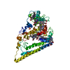
| Structure viewer | Molecule: MolmilJmol/JSmol |
|---|
- Downloads & links
-Download
| PDBx/mmCIF format | 3dsh.cif.gz | 63.9 KB | Display | PDBx/mmCIF format |
|---|---|---|---|---|
| PDB format | pdb3dsh.ent.gz | 46.5 KB | Display | PDB format |
| PDBx/mmJSON format | 3dsh.json.gz | Tree view | PDBx/mmJSON format | |
| Others |  Other downloads Other downloads |
-Validation report
| Arichive directory | https://data.pdbj.org/pub/pdb/validation_reports/ds/3dshftp://data.pdbj.org/pub/pdb/validation_reports/ds/3dsh | HTTPS FTP |
|---|
-Related structure data
| Similar structure data |
|---|


-Links
 PDBj
PDBj

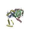
- Assembly
| Deposited unit | 
| ||||||||
|---|---|---|---|---|---|---|---|---|---|
| 1 | 
| ||||||||
| Unit cell |
|
-Components
| #1: Protein | Interferon regulatory factors / IRF-5 Mass: 28595.842 Da / Num. of mol.: 1 Fragment: IRF-5 transactivation domain (UNP residues 232-477) Mutation: S430D Source method: isolated from a genetically manipulated source Source: (gene. exp.) Homo sapiens (human) / Gene: IRF5 / Plasmid: pGEX-6p-1 / Production host:  Escherichia coli (E. coli) / Strain (production host): HB101 / References: UniProt: Q13568 Escherichia coli (E. coli) / Strain (production host): HB101 / References: UniProt: Q13568 |
|---|---|
| #2: Water | ChemComp-HOH / Water Mass: 18.015 Da / Num. of mol.: 132 / Source method: isolated from a natural source / Formula: H2O Mass: 18.015 Da / Num. of mol.: 132 / Source method: isolated from a natural source / Formula: H2O |
-Experimental details
-Experiment
| Experiment | Method: X-RAY DIFFRACTION / Number of used crystals: 1 |
|---|
- Sample preparation
| Crystal | Density Matthews: 3.45 Å3/Da / Density % sol: 64.37 % |
|---|---|
| Crystal grow | Temperature: 298 K / Method: vapor diffusion, hanging drop / pH: 7 Details: 0.5-2.0% PEG 6000, 100mM imidazole, pH 7.0, VAPOR DIFFUSION, HANGING DROP, temperature 298K |
-Data collection
| Diffraction |
| ||||||||||||||||||
|---|---|---|---|---|---|---|---|---|---|---|---|---|---|---|---|---|---|---|---|
| Diffraction source |
| ||||||||||||||||||
| Detector |
| ||||||||||||||||||
| Radiation |
| ||||||||||||||||||
| Radiation wavelength |
| ||||||||||||||||||
| Reflection | Resolution: 2→100 Å / Num. all: 26875 / Num. obs: 26875 / % possible obs: 96.9 % / Observed criterion σ(F): 0 / Observed criterion σ(I): 0 / Rmerge(I) obs: 0.057 / Net I/σ(I): 13.8 | ||||||||||||||||||
| Reflection shell | Resolution: 2→2.07 Å / Redundancy: 7.2 % / Rmerge(I) obs: 0.0229 / % possible all: 78.8 |

- Processing
| Software |
| ||||||||||||||||||||||||||||||||||||||||||||||||||||||||||||||||||||||||||||||||||||||||||||||||||||||||||||||||||||||||||||||||||||||||||||||||||||||||||||||||||||||||||
|---|---|---|---|---|---|---|---|---|---|---|---|---|---|---|---|---|---|---|---|---|---|---|---|---|---|---|---|---|---|---|---|---|---|---|---|---|---|---|---|---|---|---|---|---|---|---|---|---|---|---|---|---|---|---|---|---|---|---|---|---|---|---|---|---|---|---|---|---|---|---|---|---|---|---|---|---|---|---|---|---|---|---|---|---|---|---|---|---|---|---|---|---|---|---|---|---|---|---|---|---|---|---|---|---|---|---|---|---|---|---|---|---|---|---|---|---|---|---|---|---|---|---|---|---|---|---|---|---|---|---|---|---|---|---|---|---|---|---|---|---|---|---|---|---|---|---|---|---|---|---|---|---|---|---|---|---|---|---|---|---|---|---|---|---|---|---|---|---|---|---|---|
| Refinement | Method to determine structure: MIR / Resolution: 2→30.46 Å / Cor.coef. Fo:Fc: 0.955 / Cor.coef. Fo:Fc free: 0.938 / SU B: 8.285 / SU ML: 0.104 / TLS residual ADP flag: LIKELY RESIDUAL / Cross valid method: THROUGHOUT / σ(F): 0 / ESU R: 0.146 / ESU R Free: 0.142 / Stereochemistry target values: MAXIMUM LIKELIHOOD
| ||||||||||||||||||||||||||||||||||||||||||||||||||||||||||||||||||||||||||||||||||||||||||||||||||||||||||||||||||||||||||||||||||||||||||||||||||||||||||||||||||||||||||
| Solvent computation | Ion probe radii: 0.8 Å / Shrinkage radii: 0.8 Å / VDW probe radii: 1.2 Å / Solvent model: MASK | ||||||||||||||||||||||||||||||||||||||||||||||||||||||||||||||||||||||||||||||||||||||||||||||||||||||||||||||||||||||||||||||||||||||||||||||||||||||||||||||||||||||||||
| Displacement parameters | Biso mean: 45.605 Å2
| ||||||||||||||||||||||||||||||||||||||||||||||||||||||||||||||||||||||||||||||||||||||||||||||||||||||||||||||||||||||||||||||||||||||||||||||||||||||||||||||||||||||||||
| Refinement step | Cycle: LAST / Resolution: 2→30.46 Å
| ||||||||||||||||||||||||||||||||||||||||||||||||||||||||||||||||||||||||||||||||||||||||||||||||||||||||||||||||||||||||||||||||||||||||||||||||||||||||||||||||||||||||||
| Refine LS restraints |
| ||||||||||||||||||||||||||||||||||||||||||||||||||||||||||||||||||||||||||||||||||||||||||||||||||||||||||||||||||||||||||||||||||||||||||||||||||||||||||||||||||||||||||
| LS refinement shell | Resolution: 1.998→2.05 Å / Total num. of bins used: 20
| ||||||||||||||||||||||||||||||||||||||||||||||||||||||||||||||||||||||||||||||||||||||||||||||||||||||||||||||||||||||||||||||||||||||||||||||||||||||||||||||||||||||||||
| Refinement TLS params. | Method: refined / Origin x: 11.439 Å / Origin y: 35.825 Å / Origin z: 72.601 Å
|
