| Entry | Database: PDB / ID: 4rwt
|
|---|
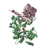
| Title | Structure of actin-Lmod complex |
|---|
 Components Components | |
|---|
 Keywords Keywords |  STRUCTURAL PROTEIN / leucine rich region / actin nucleation / actin STRUCTURAL PROTEIN / leucine rich region / actin nucleation / actin |
|---|
| Function / homology |  Function and homology information Function and homology information
Gap junction degradation / Formation of annular gap junctions / EPHB-mediated forward signaling / EPH-ephrin mediated repulsion of cells / Recycling pathway of L1 / VEGFA-VEGFR2 Pathway / : / Cell-extracellular matrix interactions / RHOBTB2 GTPase cycle / RHOF GTPase cycle ...Gap junction degradation / Formation of annular gap junctions / EPHB-mediated forward signaling / EPH-ephrin mediated repulsion of cells / Recycling pathway of L1 / VEGFA-VEGFR2 Pathway / : / Cell-extracellular matrix interactions / RHOBTB2 GTPase cycle / RHOF GTPase cycle / pointed-end actin filament capping / MAP2K and MAPK activation / Platelet degranulation / RHO GTPases Activate WASPs and WAVEs / Regulation of actin dynamics for phagocytic cup formation / DNA Damage Recognition in GG-NER / UCH proteinases / Clathrin-mediated endocytosis / myofibril assembly / sperm individualization / actin nucleation / brahma complex / maintenance of protein location in cell / tube formation / Ino80 complex / M band / sarcomere organization / tropomyosin binding / myofibril / positive regulation of actin filament polymerization / striated muscle thin filament / mitotic cytokinesis / actin monomer binding / actin filament polymerization / sarcomere / muscle contraction / actin filament organization / actin filament / Hydrolases; Acting on acid anhydrides; Acting on acid anhydrides to facilitate cellular and subcellular movement / actin binding / cytoskeleton / hydrolase activity / chromatin remodeling / ATP binding / cytoplasmSimilarity search - Function Tropomodulin / Tropomodulin / WH2 domain / WH2 domain profile. / Leucine-rich repeat, LRR (right-handed beta-alpha superhelix) / Ribonuclease Inhibitor / ATPase, substrate binding domain, subdomain 4 / Actin; Chain A, domain 4 / Alpha-Beta Horseshoe / ATPase, nucleotide binding domain ...Tropomodulin / Tropomodulin / WH2 domain / WH2 domain profile. / Leucine-rich repeat, LRR (right-handed beta-alpha superhelix) / Ribonuclease Inhibitor / ATPase, substrate binding domain, subdomain 4 / Actin; Chain A, domain 4 / Alpha-Beta Horseshoe / ATPase, nucleotide binding domain / Actins signature 1. / Actin, conserved site / Actins signature 2. / Actin/actin-like conserved site / Actins and actin-related proteins signature. / Actin / Actin family / Actin / Leucine-rich repeat domain superfamily / ATPase, nucleotide binding domain / Nucleotidyltransferase; domain 5 / Alpha-Beta Complex / 2-Layer Sandwich / Alpha BetaSimilarity search - Domain/homology |
|---|
| Biological species |   Drosophila melanogaster (fruit fly) Drosophila melanogaster (fruit fly)
 Homo sapiens (human) Homo sapiens (human) |
|---|
| Method | X-RAY DIFFRACTION / SYNCHROTRON / MOLECULAR REPLACEMENT / Resolution: 2.98 Å |
|---|
 Authors Authors | Chen, X. / Ni, F. / Wang, Q. |
|---|
 Citation Citation | Journal: Proc.Natl.Acad.Sci.USA / Year: 2015
Title: Mechanisms of leiomodin 2-mediated regulation of actin filament in muscle cells.
Authors: Chen, X. / Ni, F. / Kondrashkina, E. / Ma, J. / Wang, Q. |
|---|
| History | | Deposition | Dec 5, 2014 | Deposition site: RCSB / Processing site: RCSB |
|---|
| Revision 1.0 | Oct 14, 2015 | Provider: repository / Type: Initial release |
|---|
| Revision 1.1 | Oct 28, 2015 | Group: Database references |
|---|
| Revision 1.2 | Feb 28, 2024 | Group: Data collection / Database references / Derived calculations
Category: chem_comp_atom / chem_comp_bond ...chem_comp_atom / chem_comp_bond / database_2 / pdbx_struct_conn_angle / struct_conn / struct_ref_seq_dif / struct_site
Item: _database_2.pdbx_DOI / _database_2.pdbx_database_accession ..._database_2.pdbx_DOI / _database_2.pdbx_database_accession / _pdbx_struct_conn_angle.ptnr1_auth_asym_id / _pdbx_struct_conn_angle.ptnr1_auth_comp_id / _pdbx_struct_conn_angle.ptnr1_auth_seq_id / _pdbx_struct_conn_angle.ptnr1_label_asym_id / _pdbx_struct_conn_angle.ptnr1_label_atom_id / _pdbx_struct_conn_angle.ptnr1_label_comp_id / _pdbx_struct_conn_angle.ptnr1_label_seq_id / _pdbx_struct_conn_angle.ptnr2_auth_asym_id / _pdbx_struct_conn_angle.ptnr2_label_asym_id / _pdbx_struct_conn_angle.ptnr3_auth_asym_id / _pdbx_struct_conn_angle.ptnr3_auth_comp_id / _pdbx_struct_conn_angle.ptnr3_auth_seq_id / _pdbx_struct_conn_angle.ptnr3_label_asym_id / _pdbx_struct_conn_angle.ptnr3_label_atom_id / _pdbx_struct_conn_angle.ptnr3_label_comp_id / _pdbx_struct_conn_angle.ptnr3_label_seq_id / _pdbx_struct_conn_angle.value / _struct_conn.pdbx_dist_value / _struct_conn.ptnr1_auth_asym_id / _struct_conn.ptnr1_auth_comp_id / _struct_conn.ptnr1_auth_seq_id / _struct_conn.ptnr1_label_asym_id / _struct_conn.ptnr1_label_atom_id / _struct_conn.ptnr1_label_comp_id / _struct_conn.ptnr1_label_seq_id / _struct_conn.ptnr2_auth_asym_id / _struct_conn.ptnr2_label_asym_id / _struct_ref_seq_dif.details / _struct_site.pdbx_auth_asym_id / _struct_site.pdbx_auth_comp_id / _struct_site.pdbx_auth_seq_id |
|---|
|
|---|
 Movie
Movie Controller


 Open data
 Basic information
 Structure visualization
 Downloads & links
 Other downloads


 PDBj
PDBj


 Assembly
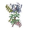


 Mass: 506.196 Da / Num. of mol.: 2 / Source method: obtained synthetically / Formula: C10H17N6O12P3 / Comment: AMP-PNP, energy-carrying molecule analogue*YM
 Mass: 24.305 Da / Num. of mol.: 2 / Source method: obtained synthetically / Formula: Mg
 Sample preparation
 / Beamline: 21-ID-F / Wavelength: 0.97872 Å
 Processing